


 Inglés (pdf)
 Articulo en XML
 Referencias del artículo
 Enviar articulo por email
 Citado por SciELO
  Similares en
SciELO
Similares en
SciELO 
 Permalink
A previously healthy 23-year-old male patient presented with a brown spot on his forehead, first noticed at the age of 13, with no reported changes in the previous years. A sharply well-circumscribed 60 × 30 mm light brown patch on the right forehead with no hypertrichosis was noted (Fig. 1A). There was no firmness, palpable mass, or tenderness on palpation. Dermoscopy revealed a homogeneous melanocytic lesion with a faint reticular pattern and perifollicular hypopigmentation (Figs. 1B and 1C). Histopathological examination showed acanthosis and increased pigmentation in the basal layer of the epidermis. Based on clinical, dermoscopic, and histological findings, Becker's melanosis was diagnosed. Laser therapy was offered; however, the patient refused. Becker's melanosis, also known as Becker's nevus, is a relatively common benign cutaneous hamartoma with epidermal or dermal elements1. It is clinically characterized by well-circumscribed, unilateral, acquired hyperpigmentation, usually first noticed around puberty and more prevalent in males2. Although it is most frequently seen in the shoulder, scapular area, and upper extremity, it can be seen anywhere2. When Becker melanosis is referred to as Becker nevus syndrome, it may occasionally be linked to developmental abnormalities like ipsilateral breast hypoplasia, extra nipples, aplasia of the pectoralis major muscle, and other musculoskeletal and spine abnormalities3. The differential diagnosis of Becker melanosis includes congenital melanocytic nevus, congenital smooth muscle hamartoma, plexiform neurofibroma, post-inflammatory hyperpigmentation, and café-au-lait macules2,4. Diagnosis is mainly clinical; however, a skin biopsy can be helpful to support the diagnosis of Becker's melanosis, mainly when dealing with atypical presentations.
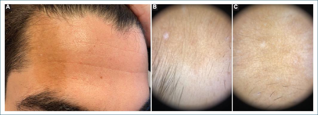
Figure 1 A: a sharply circumscribed 60 × 30 mm light brown patch on the right forehead. B and C: dermoscopy (dermlite DL4 polarized mode) showed a faint reticular pattern of pigmentation, perifollicular hypopigmentation, and a sharply demarcated unstructured white area (biopsy site).
Facial Becker nevus is not widespread and hypertrichosis does not always accompany it. There is a need for larger studies on whether this case is associated with any anomaly and its prevalence. The purpose of reporting our case is to remind and emphasize that Becker's nevus may be outside of the usual areas and should be kept in mind in the differential diagnosis of hyperpigmented, sharply demarcated spots on the face.

