<?xml version="1.0" encoding="ISO-8859-1"?><article xmlns:mml="http://www.w3.org/1998/Math/MathML" xmlns:xlink="http://www.w3.org/1999/xlink" xmlns:xsi="http://www.w3.org/2001/XMLSchema-instance">
<front>
<journal-meta>
<journal-id>0872-0754</journal-id>
<journal-title><![CDATA[Nascer e Crescer]]></journal-title>
<abbrev-journal-title><![CDATA[Nascer e Crescer]]></abbrev-journal-title>
<issn>0872-0754</issn>
<publisher>
<publisher-name><![CDATA[Centro Hospitalar do Porto]]></publisher-name>
</publisher>
</journal-meta>
<article-meta>
<article-id>S0872-07542018000100012</article-id>
<title-group>
<article-title xml:lang="en"><![CDATA[Imaging case]]></article-title>
<article-title xml:lang="pt"><![CDATA[Caso imagiológico]]></article-title>
</title-group>
<contrib-group>
<contrib contrib-type="author">
<name>
<surname><![CDATA[Rangel]]></surname>
<given-names><![CDATA[Maria Adriana]]></given-names>
</name>
<xref ref-type="aff" rid="A01"/>
</contrib>
<contrib contrib-type="author">
<name>
<surname><![CDATA[Loureiro]]></surname>
<given-names><![CDATA[Graça]]></given-names>
</name>
<xref ref-type="aff" rid="A02"/>
</contrib>
<contrib contrib-type="author">
<name>
<surname><![CDATA[Pinto]]></surname>
<given-names><![CDATA[Mariana]]></given-names>
</name>
<xref ref-type="aff" rid="A02"/>
</contrib>
</contrib-group>
<aff id="A01">
<institution><![CDATA[,Centro Hospitalar de Vila Nova de Gaia e Espinho Department of Pediatrics ]]></institution>
<addr-line><![CDATA[Vila Nova de Gaia ]]></addr-line>
<country>Portugal</country>
</aff>
<aff id="A02">
<institution><![CDATA[,Centro Hospitalar Entre Douro e Vouga Department of Pediatrics ]]></institution>
<addr-line><![CDATA[Santa Maria da Feira ]]></addr-line>
<country>Portugal</country>
</aff>
<pub-date pub-type="pub">
<day>01</day>
<month>03</month>
<year>2018</year>
</pub-date>
<pub-date pub-type="epub">
<day>01</day>
<month>03</month>
<year>2018</year>
</pub-date>
<volume>27</volume>
<numero>1</numero>
<fpage>61</fpage>
<lpage>64</lpage>
<copyright-statement/>
<copyright-year/>
<self-uri xlink:href="http://scielo.pt/scielo.php?script=sci_arttext&amp;pid=S0872-07542018000100012&amp;lng=en&amp;nrm=iso"></self-uri><self-uri xlink:href="http://scielo.pt/scielo.php?script=sci_abstract&amp;pid=S0872-07542018000100012&amp;lng=en&amp;nrm=iso"></self-uri><self-uri xlink:href="http://scielo.pt/scielo.php?script=sci_pdf&amp;pid=S0872-07542018000100012&amp;lng=en&amp;nrm=iso"></self-uri><abstract abstract-type="short" xml:lang="en"><p><![CDATA[Introduction: Cephalohematoma is a collection of serosanguineous fluid below the periosteum and is the most frequent cranial injury in the newborn, occurring in 0.2-2.5% live births. The majority of cephalohematomas spontaneously resolve within three to four weeks, however, some persist beyond four weeks and begin to calcify. Case report: A seven-week-old boy, was referred to the emergency department because of a head lump on the right parietal region, with no other symptoms. He was born after a vacuum-assisted delivery, and presented a cephalohematoma in the first days of life, that progressively decreased and became more rigid. Physical examination, revealed a cranial asymmetry, and a head lump on the right parietal region, that was hard and fixed to the bone. Head X-ray revealed a radiopaque lump on the right parietal bone and a poorly defined arched line, as well as visible microcalcifications on the core of the cephalohematoma, typical findings of a calcified cephalohematoma. Discussion: Even though cephalohematoma is frequently encountered, calcified cephalohematoma is seen only sporadically, and is a rare clinical entity. History and clinical examination are important in the differential diagnosis and imaging strategy. Radiography and ultrasonography are often the initial screening diagnostic tests, followed by magnetic resonance imaging or computed tomography. Head x-ray features, in this case report, where particularly evocative of the diagnosis.]]></p></abstract>
<abstract abstract-type="short" xml:lang="pt"><p><![CDATA[Introdução: O cephalohematoma é uma coleção sero-hemática subperióssea e é o traumatismo craniano mais frequente do recém-nascido, ocorrendo em 0,2-2,5% dos nados vivos. A maioria sofre regressão espontânea pelas três a quatro semanas de vida, sendo que uma minoria persiste após as quatro semanas, podendo evoluir para calcificação. Caso Clínico: Lactente de sete semanas, sexo masculino, referenciado ao serviço de urgência por tumefação craniana na região parietal direita. Nascido de parto distócico por ventosa; observou-se cefalohematoma nos primeiros dias de vida, o qual diminuiu progressivamente de tamanho, tornando-se mais rígido. Sem qualquer outra sintomatologia. Ao exame físico era notória, uma assimetria craniana e uma tumefação na região parietal direita, dura e fixa à palpação do crânio. A radiografia de crânio revelou uma tumefação radiopaca parietal direita, associada a uma linha arqueada mal definida, bem como microcalcificações na parte central do cefalohematoma, achados típicos de um cefalohematoma calcificado. Conclusão: Embora o cefalohematoma seja frequentemente observado, o cefalohematoma calcificado é visto apenas esporadicamente e é uma entidade clínica rara. A história clinica e exame físico são importantes para o diagnóstico diferencial e planificação do estudo imagiológico. A radiografia e a ecografia são habitualmente os exames de primeira linha seguidas pela ressonância magnética ou tomografia computorizada. As características da radiografia de crânio, neste caso clínico, foram particularmente sugestivas do diagnóstico.]]></p></abstract>
<kwd-group>
<kwd lng="en"><![CDATA[Birth Injuries]]></kwd>
<kwd lng="en"><![CDATA[calcified cephalohematoma]]></kwd>
<kwd lng="en"><![CDATA[newborn]]></kwd>
<kwd lng="pt"><![CDATA[Cefalohematoma calcificado]]></kwd>
<kwd lng="pt"><![CDATA[recém-nascido]]></kwd>
<kwd lng="pt"><![CDATA[traumatismos do nascimento]]></kwd>
</kwd-group>
</article-meta>
</front><body><![CDATA[  <font face="Verdana" size="2">     <p align="right"><b>QUAL O SEU DIAGN&Oacute;STICO? | WHAT IS YOUR DIAGNOSIS?</b></p>     <p>&nbsp;</p> </font>     <p><font size="4" face="Verdana"><b>Imaging case</b></font></p> <font face="Verdana" size="2">     <p>&nbsp;</p> </font>     <p><font size="3" face="Verdana"><b>Caso imagiol&oacute;gico</b></font></p>     <p>&nbsp;</p>     <p>&nbsp;</p> <font face="Verdana" size="2">     <p><b>Maria Adriana Rangel<sup>I</sup>, Graça Loureiro<sup>II</sup>, Mariana   Pinto<sup>II</sup></b></p>     <p><sup>I </sup>Department of Pediatrics,   Centro Hospitalar de Vila Nova de Gaia e Espinho. 4400-129 Vila Nova de Gaia, Portugal. <a href="mailto:mariaadrianarangel@hotmail.com">mariaadrianarangel@hotmail.com</a>    ]]></body>
<body><![CDATA[<br> <sup>II </sup>Department of Pediatrics, Centro Hospitalar Entre Douro e Vouga. 4520-161 Santa Maria da Feira, Portugal. <a href="mailto:mariagracaloureiro@gmail.com">mariagracaloureiro@gmail.com</a>; <a href="mailto:marianabrpinto@gmail.com">marianabrpinto@gmail.com</a></p> <a href="#end">Correspondence to</a><a name="topo" id="topo"></a>     <p>&nbsp;</p>     <p>&nbsp;</p> </font> <hr noshade size="1"> <font face="Verdana" size="2">     <p><b>ABSTRACT</b></p>     <p><b>Introduction:</b> Cephalohematoma is a collection of serosanguineous fluid below the   periosteum and is the most frequent cranial injury in the newborn, occurring in   0.2-2.5% live births. The majority of cephalohematomas spontaneously resolve   within three to four weeks, however, some persist beyond four weeks and begin to calcify.</p>     <p><b>Case report</b>: A seven-week-old boy, was referred to the emergency department because   of a head lump on the right parietal region, with no other symptoms. He was   born after a vacuum-assisted delivery, and presented a cephalohematoma in the   first days of life, that progressively decreased and became more rigid.   Physical examination, revealed a cranial asymmetry, and a head lump on the   right parietal region, that was hard and fixed to the bone. Head X-ray revealed   a radiopaque lump on the right parietal bone and a poorly defined arched line,   as well as visible microcalcifications on the core of the cephalohematoma, typical findings of a calcified cephalohematoma. </p>     <p><b>Discussion:</b> Even though cephalohematoma is frequently encountered, calcified   cephalohematoma is seen only sporadically, and is a rare clinical entity.   History and clinical examination are important in the differential diagnosis   and imaging strategy. Radiography and ultrasonography are often the initial   screening diagnostic tests, followed by magnetic resonance imaging or computed   tomography. Head x-ray features, in this case report, where particularly evocative of the diagnosis.</p>     <p><b>Keywords:</b> Birth Injuries; calcified cephalohematoma; newborn</p> </font> <hr noshade size="1"> <font face="Verdana" size="2">     <p><b>RESUMO</b></p>     <p><b>Introdução:</b> O cephalohematoma é uma coleção sero-hemática subperióssea e é o   traumatismo craniano mais frequente do recém-nascido, ocorrendo em 0,2-2,5% dos   nados vivos. A maioria sofre regressão espontânea pelas três a quatro semanas   de vida, sendo que uma minoria persiste após as quatro semanas, podendo evoluir para calcificação. </p>     ]]></body>
<body><![CDATA[<p><b>Caso Clínico:</b> Lactente de sete semanas, sexo masculino, referenciado ao serviço de   urgência por tumefação craniana na região parietal direita. Nascido de parto   distócico por ventosa; observou-se cefalohematoma nos primeiros dias de vida, o   qual diminuiu progressivamente de tamanho, tornando-se mais rígido. Sem   qualquer outra sintomatologia.  Ao exame   físico era notória, uma assimetria craniana e uma tumefação na região parietal direita,   dura e fixa à palpação do crânio. A radiografia de crânio revelou uma tumefação   radiopaca parietal direita, associada a uma linha arqueada mal definida, bem   como microcalcificações na parte central do cefalohematoma, achados típicos de um cefalohematoma calcificado. </p>     <p><b>Conclusão:</b> Embora o   cefalohematoma seja frequentemente observado, o cefalohematoma calcificado é   visto apenas esporadicamente e é uma entidade clínica rara. A história clinica   e exame físico são importantes para o diagnóstico diferencial e planificação do   estudo imagiológico. A radiografia e a ecografia são habitualmente os exames de   primeira linha seguidas pela ressonância magnética ou tomografia computorizada.   As características da radiografia de crânio, neste caso clínico, foram particularmente sugestivas do diagnóstico.</p>     <p><b>Palavras-chave:</b> Cefalohematoma calcificado; recém-nascido; traumatismos do nascimento</p> </font> <hr noshade size="1"> <font face="Verdana" size="2">     <p>&nbsp;</p>     <p>&nbsp;</p>     <p>A seven-week-old boy was referred for presenting   a hard mass over the right parietal region of the skull.  A soft swelling was present in the first few   days of life, that progressively decreased in size and became more rigid. No   other symptoms were reported, namely irritability, vomiting or fever. The baby   was the first born of non-consanguineous healthy parents. Pregnancy was   monitored and uneventful and the baby was born at 38 weeks through   vacuum-assisted delivery, with an Apgar score of 9/10 by the 1st/5th minute. He   presented a cephalohematoma, localized at the region of attachment of the   suction cup. Neonatal period was otherwise uneventful. There was no history of   trauma besides the cephalohematoma at birth or other pathological process such as bleeding disorders. </p>     <p>
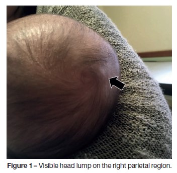
On physical examination he was calm, with appropriate toning and reflex.   A cranial asymmetry was notorious, with a visible head lump (<a href="#f1">figure 1</a>) on the   right parietal region, hard and fixed to the bone. There was no associated soft   tissue abnormality, and the margins were indistinct. The scalp was freely   moving over the swelling and apparently painless, since there was no grimace of pain or crying during palpation. Remaining physical examination unremarkable.
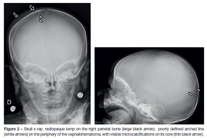
 </p>     <p><a name="f1"></a></p>     <p>&nbsp;</p>     <p align="center"><img src="/img/revistas/nas/v27n1/27n1a12f1.jpg" width="352" height="344"></p>     
]]></body>
<body><![CDATA[<p>&nbsp;</p>     <p>A head X-ray (<a href="/img/revistas/nas/v27n1/27n1a12f2.jpg">figure 2</a>) revealed a radiopaque lump on the right parietal   bone (large black arrow) and a poorly defined arched line (white arrows) on the   periphery of the cephalohematoma, with visible microcalcifications on its core (thin black arrow). No cranial fracture was seen.</p>     
<p><b>What is your diagnosis?</b></p>     <p>&nbsp;</p> </font>     <p><font size="3" face="Verdana"><b>DIAGNOSIS</b></font></p> <font face="Verdana" size="2">     <p>Calcified cephalohematoma (CC)</p>     <p>&nbsp;</p> </font>     <p><font size="3" face="Verdana"><b>DISCUSSION</b></font></p> <font face="Verdana" size="2">     <p>Birth injuries to the head occur because the head is particularly   vulnerable to trauma during vertex delivery.<sup>1</sup> Cephalohematoma is the   most frequent cranial injury in the newborn, occurring in 0.2-2.5% live births,   with parietal bone being the most common site of involvement.<sup>2,3</sup>   However there are reports of diagnosis <i>in útero</i> by prenatal ultrasound or after cesarean section.<sup>1</sup> </p>     <p>Cephalohematoma is a collection of serosanguineous or bloody fluid below   the periosteum of the skull. It results from bleeding of the emissary and   diploic veins into the subperiostial layer. The bleeding is contained by the   ligaments that attach the periosteum to the skull at the cranial suture lines.   The pressure in this area builds and acts as a tamponade to stop further   bleeding. However, as it occurs in a deeper and more vascular portion of the scalp, it usually has a higher content of blood than caput succedaneum.<sup>1</sup></p>     ]]></body>
<body><![CDATA[<p>Clinically, cephalohematoma is not usually   present at birth but develop hours or even days after delivery, as an immobile,   firm, localized mass with cranial sutures clearly defining the boundaries.<sup>1,4</sup>   It occurs more frequently in <i>primigravidas</i>, in male gender or macrosomic   newborns, in instrumented assisted deliveries (vacuum or forceps), prolonged   difficult labour, cephalopelvic disproportion, head in deviant position (occipital transverse) or when a scalp electrode has been placed.<sup>1</sup></p>     <p>Complications of cephalohematoma may include: anaemia (albeit  rare) and hyperbilirubinemia (due to   resolving hematoma); underlined linear skull fractures (5-20%); or rarely meningitis, osteomyelitis or intracranial injury.<sup>4</sup></p>     <p>The majority of cephalohematomas spontaneously resorb, by gradual   haemolysis; the swelling becomes increasingly fluctuant and generally resolves   in three to four weeks.<sup>3,4</sup> In about 3-5% of cases, cephalohematoma   persist beyond four weeks and progresses to calcification.<sup>2,3</sup> This   results from a process of subperiostial osteogenesis and calcium deposits in   the area. The calcification process begins when a ridge forms along the   periphery of the hematoma, causing distortion of the calvaria.<sup>2</sup> The   rate of sub-pericranium osteogenesis is variable.<sup>3</sup> If sufficiently   large, the hematoma can depress the malleable neonatal skull, causing it to   encroach into the cranial vault space as the cephalohematoma expands. This   supports the current classification of Calcified Cephalohematoma (CC) in two   types: type 1, non-depressed with no encroachment into the cranial cavity; and   type 2, depression of the inner bone and encroachment. This classification is   important as it determines the appropriate corrective surgical technique.<sup>3 </sup></p>     <p>Although rare, calcified or ossified cephalohematomas can cause   significant deformities to the skull requiring treatment. Besides the aesthetic   effects, clinical sequelae are rarely reported (even in type 2 CC), however,   electroencephalography anomalies have been reported, and association with   sagittal synostosis, although this is not clear and still subject of debate.<sup>2,5</sup>   While there are currently no reported cases of CC causing focal neurological   deficits or disturbances in intracranial pressures, the long-term effects of a   calcified mass encroaching into the cranial vault (in type 2 CC) on brain development is unknown.<sup>3</sup></p>     <p>Neuroimaging is the main investigation needed for CC. Duplex/color   doppler ultrasound (US) in the evaluation of head bumps and lumps provides   rapid acquisition of information including size, shape, location, internal   content and vascularity.<sup>6,7</sup> This imaging technique does not expose   patients to radiation or iodinated contrast nor requires sedation. Skull X-ray   features includes an expansible lesion surrounded by a radio-opaque rim, as   seen in this case (<a href="/img/revistas/nas/v27n1/27n1a12f2.jpg">figure 2</a>).<sup>3</sup> There may be a variable thinning of   the underlying calvarium.<sup>3</sup> CT-Neuroimaging may be needed to exclude   a deformation of the inner carinal surface, for assessment of type of lesion   and for operative planning. It may also be useful for differential diagnosis   such as bone tumour or scalloping of skull due to brain tumours/lesions and to   exclude an intracranial hematoma.<sup>2</sup> Despite the use of radiation, CT   is the standard for assessment of CC.<sup>7 </sup>Its features include a uniformly   homogenous, hypodense, non-enhancing core encased by bone.<sup>3</sup>   Three-dimensional reconstructive CT allows preoperative planning of appropriate   reconstructive technique.2 On Magnetic Resonance Imaging (MRI), cephalohematoma   exhibits a bright signal on T1-weighted images and a predominantly high signal   with mild heterogeneity on T2-weighted images, due to the presence of   methaemoglobin, indicating that the lesion is secondary to haemorrhage rather than to an intraosseous tumor.<sup>2,3</sup></p>     
<p>After ossification, cephalohematoma may undergo   spontaneous remodelling and disappear within   three to six months. Persistent calcification, significant deformity and/or secondary cranial complications     may be an indication for surgical excision.  <sup>4,8,9</sup> Treatment of   CC is still controversial due to the possibility of a benign clinical course.   In neonates, the approach is usually conservative, because the cephalohematoma   can be gradually absorbed as the skull develops. Some authors recommend an   aspiration attempt for any significantly sized cephalohematoma that fail to   reabsorb after a month.<sup>3</sup> However, the benefit of aspiration must be   outweighed with the risk of infection.<sup>4</sup> A passive cranial moulding   helmet therapy as a successful nonsurgical treatment for CC causing cranial   asymmetry has also been proposed. Moulding is only effective in partially   calcified cephalohematomas and probably would yield poor results in completely   CC. Furthermore, while moulding is effective in reshaping the skull, this   process may push the inner lamella of the cephalohematoma further into the   cranial vault, potentially converting a Type 1 into a Type 2 lesion.<sup>3</sup> The risk of recurrence with this treatment is also unknown.</p>     <p>A relatively consensual indication for surgery is the correction of   skull asymmetry for cosmetic reasons.<sup>3,9 </sup>Other indications may be   the prevention of brain growth restriction, diagnostic confirmation and   treatment of associated craniosynostosis.<sup>2,9</sup> While experience with   surgical correction of CC is limited, the safety and efficacy of intracranial   surgery for correction of skull asymmetry in other conditions such as   craniosynostosis in children have been well documented, and therefore surgical correction for large calcified cephalohematomas should be considered.<sup>3</sup></p>     <p>&nbsp;</p> </font>     <p><font size="3" face="Verdana"><b>CONCLUSION</b></font></p> <font face="Verdana" size="2">     <p>Even though cephalohematoma is frequently encountered, calcified   cephalohematoma is seen only sporadically, and is a rare clinical entity. The   history and clinical examination are important in the differential diagnosis   and imaging strategy. Radiography and ultrasonography are often the initial   screening diagnostic tests, followed by magnetic resonance imaging or computed   tomography. Head x-ray features, in this case report, where particularly evocative of the diagnosis.</p>      ]]></body>
<body><![CDATA[<p>&nbsp;</p> </font>     <p><font size="3" face="Verdana"><b>REFERENCES</b></font></p> <font face="Verdana" size="2"> <ol>       <li>         <p>Nicholson       L. Caput succedaneum and cephalohematoma: the cs that leave bumps on the head.       Neonatal Netw. 2007; 26:277-81.</p>   </li>       <li>         <p>Vigo       V, Battaglia DI, Frassanito P, Tamburrini G, Caldarelli M, Massimi L. Calcified       cephalohematoma as an unusual cause of EEG anomalies: case report. J Neurosurg       Pediatr. 2017; 19:46-50.</p>   </li>       <li>         <p>Wong       CH, Foo CL, Seow WT. Calcified cephalohematoma: classification, indications for       surgery and techniques. J Craniofac Surg. 2006; 17:970-9.</p>   </li>       <li>         <p>Guclu       B, Yalcinkaya U, Kazanci B, Adilay U, Ekici MA. Diagnosis and treatment of ossified       cephalhematoma. J Craniofac Surg. 2012; 23:e505-7.</p>   </li>       ]]></body>
<body><![CDATA[<li>         <p>Piatt       JH, Jr. Sagittal synostosis and ossified scalp hematoma: cause or consequence?       J Neurosurg Pediatr. 2010; 6:29-32.</p>   </li>       <li>         <p>Bansal       AG, Rosenberg HK. Sonography of pediatric superficial lumps and bumps:       illustrative examples from head to toe. Pediatr Radiol. 2017; 47:1171-83.</p>   </li>       <li>         <p>Moron       FE, Morriss MC, Jones JJ, Hunter JV. Lumps and bumps on the head in children:       use of CT and MR imaging in solving the clinical diagnostic dilemma.       Radiographics. 2004; 24:1655-74</p>   </li>       <li>         <p>Liu       L, Dong C, Chen L. Surgical Treatment of Ossified cephalohematoma: A Case       Report and Review of the Literature. World Neurosurg. 2016; 96:614 e7- e9.</p>   </li>       <li>         <p>Krishnan       P, Karthigeyan M, Salunke P. Ossified Cephalhematoma: An Unusual Cause of Calvarial       Mass in Infancy J Pediatr Neurosci. 2017; 12: 64–66<i>. </i></p>   </li>       ]]></body>
<body><![CDATA[</ol>     <p>&nbsp;</p>     <p>&nbsp;</p> <b><a name="end" id="topo2"></a><a href="#topo">CORRESPONDENCE TO</a></b>     <p>Maria Adriana Rangel    <br> Department of Pediatrics    <br> Centro Hospitalar de Vila Nova de Gaia e Espinho    <br> Rua Dr. Francisco Sá Carneiro    <br> 4400-129 Vila Nova de Gaia    <br> Email: <a href="mailto:mariaadrianarangel@hotmail.com">mariaadrianarangel@hotmail.com</a></p>     <p>Received for publication: 05.09.2017 Accepted in revised form: 08.01.2018</p> </font>     ]]></body>
<body><![CDATA[ ]]></body><back>
<ref-list>
<ref id="B1">
<label>1</label><nlm-citation citation-type="journal">
<person-group person-group-type="author">
<name>
<surname><![CDATA[Nicholson]]></surname>
<given-names><![CDATA[L]]></given-names>
</name>
</person-group>
<article-title xml:lang="en"><![CDATA[Caput succedaneum and cephalohematoma: the cs that leave bumps on the head]]></article-title>
<source><![CDATA[Neonatal Netw]]></source>
<year>2007</year>
<volume>26</volume>
<page-range>277-81</page-range></nlm-citation>
</ref>
<ref id="B2">
<label>2</label><nlm-citation citation-type="journal">
<person-group person-group-type="author">
<name>
<surname><![CDATA[Vigo]]></surname>
<given-names><![CDATA[V]]></given-names>
</name>
<name>
<surname><![CDATA[Battaglia]]></surname>
<given-names><![CDATA[DI]]></given-names>
</name>
<name>
<surname><![CDATA[Frassanito]]></surname>
<given-names><![CDATA[P]]></given-names>
</name>
<name>
<surname><![CDATA[Tamburrini]]></surname>
<given-names><![CDATA[G]]></given-names>
</name>
<name>
<surname><![CDATA[Caldarelli]]></surname>
<given-names><![CDATA[M]]></given-names>
</name>
<name>
<surname><![CDATA[Massimi]]></surname>
<given-names><![CDATA[L]]></given-names>
</name>
</person-group>
<article-title xml:lang="en"><![CDATA[Calcified cephalohematoma as an unusual cause of EEG anomalies: case report]]></article-title>
<source><![CDATA[J Neurosurg Pediatr]]></source>
<year>2017</year>
<volume>19</volume>
<page-range>46-50</page-range></nlm-citation>
</ref>
<ref id="B3">
<label>3</label><nlm-citation citation-type="journal">
<person-group person-group-type="author">
<name>
<surname><![CDATA[Wong]]></surname>
<given-names><![CDATA[CH]]></given-names>
</name>
<name>
<surname><![CDATA[Foo]]></surname>
<given-names><![CDATA[CL]]></given-names>
</name>
<name>
<surname><![CDATA[Seow]]></surname>
<given-names><![CDATA[WT]]></given-names>
</name>
</person-group>
<article-title xml:lang="en"><![CDATA[Calcified cephalohematoma: classification, indications for surgery and techniques]]></article-title>
<source><![CDATA[J Craniofac Surg]]></source>
<year>2006</year>
<volume>17</volume>
<page-range>970-9</page-range></nlm-citation>
</ref>
<ref id="B4">
<label>4</label><nlm-citation citation-type="journal">
<person-group person-group-type="author">
<name>
<surname><![CDATA[Guclu]]></surname>
<given-names><![CDATA[B]]></given-names>
</name>
<name>
<surname><![CDATA[Yalcinkaya]]></surname>
<given-names><![CDATA[U]]></given-names>
</name>
<name>
<surname><![CDATA[Kazanci]]></surname>
<given-names><![CDATA[B]]></given-names>
</name>
<name>
<surname><![CDATA[Adilay]]></surname>
<given-names><![CDATA[U]]></given-names>
</name>
<name>
<surname><![CDATA[Ekici]]></surname>
<given-names><![CDATA[MA]]></given-names>
</name>
</person-group>
<article-title xml:lang="en"><![CDATA[Diagnosis and treatment of ossified cephalhematoma]]></article-title>
<source><![CDATA[J Craniofac Surg]]></source>
<year>2012</year>
<volume>23</volume>
<page-range>e505-7</page-range></nlm-citation>
</ref>
<ref id="B5">
<label>5</label><nlm-citation citation-type="journal">
<person-group person-group-type="author">
<name>
<surname><![CDATA[Piatt]]></surname>
<given-names><![CDATA[JH, Jr.]]></given-names>
</name>
</person-group>
<article-title xml:lang="en"><![CDATA[Sagittal synostosis and ossified scalp hematoma: cause or consequence?]]></article-title>
<source><![CDATA[J Neurosurg Pediatr]]></source>
<year>2010</year>
<volume>6</volume>
<page-range>29-32</page-range></nlm-citation>
</ref>
<ref id="B6">
<label>6</label><nlm-citation citation-type="journal">
<person-group person-group-type="author">
<name>
<surname><![CDATA[Bansal]]></surname>
<given-names><![CDATA[AG]]></given-names>
</name>
<name>
<surname><![CDATA[Rosenberg]]></surname>
<given-names><![CDATA[HK]]></given-names>
</name>
</person-group>
<article-title xml:lang="en"><![CDATA[Sonography of pediatric superficial lumps and bumps: illustrative examples from head to toe]]></article-title>
<source><![CDATA[Pediatr Radiol]]></source>
<year>2017</year>
<volume>47</volume>
<page-range>1171-83</page-range></nlm-citation>
</ref>
<ref id="B7">
<label>7</label><nlm-citation citation-type="journal">
<person-group person-group-type="author">
<name>
<surname><![CDATA[Moron]]></surname>
<given-names><![CDATA[FE]]></given-names>
</name>
<name>
<surname><![CDATA[Morriss]]></surname>
<given-names><![CDATA[MC]]></given-names>
</name>
<name>
<surname><![CDATA[Jones]]></surname>
<given-names><![CDATA[JJ]]></given-names>
</name>
<name>
<surname><![CDATA[Hunter]]></surname>
<given-names><![CDATA[JV]]></given-names>
</name>
</person-group>
<article-title xml:lang="en"><![CDATA[Lumps and bumps on the head in children: use of CT and MR imaging in solving the clinical diagnostic dilemma]]></article-title>
<source><![CDATA[Radiographics]]></source>
<year>2004</year>
<volume>24</volume>
<page-range>1655-74</page-range></nlm-citation>
</ref>
<ref id="B8">
<label>8</label><nlm-citation citation-type="journal">
<person-group person-group-type="author">
<name>
<surname><![CDATA[Liu]]></surname>
<given-names><![CDATA[L]]></given-names>
</name>
<name>
<surname><![CDATA[Dong]]></surname>
<given-names><![CDATA[C]]></given-names>
</name>
<name>
<surname><![CDATA[Chen]]></surname>
<given-names><![CDATA[L]]></given-names>
</name>
</person-group>
<article-title xml:lang="en"><![CDATA[Surgical Treatment of Ossified cephalohematoma: A Case Report and Review of the Literature]]></article-title>
<source><![CDATA[World Neurosurg]]></source>
<year>2016</year>
<volume>96</volume>
<numero>614</numero>
<issue>614</issue>
<page-range>e7- e9</page-range></nlm-citation>
</ref>
<ref id="B9">
<label>9</label><nlm-citation citation-type="journal">
<person-group person-group-type="author">
<name>
<surname><![CDATA[Krishnan]]></surname>
<given-names><![CDATA[P]]></given-names>
</name>
<name>
<surname><![CDATA[Karthigeyan]]></surname>
<given-names><![CDATA[M]]></given-names>
</name>
<name>
<surname><![CDATA[Salunke]]></surname>
<given-names><![CDATA[P]]></given-names>
</name>
</person-group>
<article-title xml:lang="en"><![CDATA[Ossified Cephalhematoma: An Unusual Cause of Calvarial Mass in Infancy]]></article-title>
<source><![CDATA[J Pediatr Neurosci]]></source>
<year>2017</year>
<volume>12</volume>
<page-range>64-66</page-range></nlm-citation>
</ref>
</ref-list>
</back>
</article>
