<?xml version="1.0" encoding="ISO-8859-1"?><article xmlns:mml="http://www.w3.org/1998/Math/MathML" xmlns:xlink="http://www.w3.org/1999/xlink" xmlns:xsi="http://www.w3.org/2001/XMLSchema-instance">
<front>
<journal-meta>
<journal-id>0872-0754</journal-id>
<journal-title><![CDATA[Nascer e Crescer]]></journal-title>
<abbrev-journal-title><![CDATA[Nascer e Crescer]]></abbrev-journal-title>
<issn>0872-0754</issn>
<publisher>
<publisher-name><![CDATA[Centro Hospitalar do Porto]]></publisher-name>
</publisher>
</journal-meta>
<article-meta>
<article-id>S0872-07542020000300007</article-id>
<article-id pub-id-type="doi">10.25753/BirthGrowthMJ.v29.i3.16655</article-id>
<title-group>
<article-title xml:lang="en"><![CDATA[Isolated myeloid sarcoma of the Tibia]]></article-title>
<article-title xml:lang="pt"><![CDATA[Sarcoma mielóide da Tibia]]></article-title>
</title-group>
<contrib-group>
<contrib contrib-type="author">
<name>
<surname><![CDATA[Serrado]]></surname>
<given-names><![CDATA[Maria Ana]]></given-names>
</name>
<xref ref-type="aff" rid="A1"/>
</contrib>
<contrib contrib-type="author">
<name>
<surname><![CDATA[Proença]]></surname>
<given-names><![CDATA[Ana Luísa]]></given-names>
</name>
<xref ref-type="aff" rid="A2"/>
</contrib>
<contrib contrib-type="author">
<name>
<surname><![CDATA[Alves]]></surname>
<given-names><![CDATA[Pedro]]></given-names>
</name>
<xref ref-type="aff" rid="A3"/>
</contrib>
</contrib-group>
<aff id="AA1">
<institution><![CDATA[,Hospital Central do Funchal Department of Radiology ]]></institution>
<addr-line><![CDATA[Funchal ]]></addr-line>
<country>Portugal</country>
</aff>
<aff id="AA2">
<institution><![CDATA[,Centro Hospitalar de Lisboa Central Hospital Curry Cabral Department of Radiology]]></institution>
<addr-line><![CDATA[Lisboa ]]></addr-line>
<country>Portugal</country>
</aff>
<aff id="AA3">
<institution><![CDATA[,Centro Hospitalar de Lisboa Central Hospital Dona Estefânia Department of Radio Pediatrics]]></institution>
<addr-line><![CDATA[Lisboa ]]></addr-line>
<country>Portugal</country>
</aff>
<pub-date pub-type="pub">
<day>00</day>
<month>09</month>
<year>2020</year>
</pub-date>
<pub-date pub-type="epub">
<day>00</day>
<month>09</month>
<year>2020</year>
</pub-date>
<volume>29</volume>
<numero>3</numero>
<fpage>153</fpage>
<lpage>156</lpage>
<copyright-statement/>
<copyright-year/>
<self-uri xlink:href="http://scielo.pt/scielo.php?script=sci_arttext&amp;pid=S0872-07542020000300007&amp;lng=en&amp;nrm=iso"></self-uri><self-uri xlink:href="http://scielo.pt/scielo.php?script=sci_abstract&amp;pid=S0872-07542020000300007&amp;lng=en&amp;nrm=iso"></self-uri><self-uri xlink:href="http://scielo.pt/scielo.php?script=sci_pdf&amp;pid=S0872-07542020000300007&amp;lng=en&amp;nrm=iso"></self-uri><abstract abstract-type="short" xml:lang="en"><p><![CDATA[Myeloid sarcoma is a rare tumor of immature myeloid cells in an extramedullary site which can be found in any part of the body. It may precede or concurrently occur with acute myeloid leukemia, chronic myeloid leukemia, or myeloproliferative disorders/myelodysplastic syndrome. Herein is reported the rare case of a child with myeloid sarcoma in the right tibia, without leukemic involvement at diagnosis. Diagnosis was challenging and several imaging modalities (radiography, computed tomography, magnetic resonance, bone scintigraphy, positron emitted tomography-computed tomography) were required. Additionally, three biopsies were necessary to make a definitive and conclusive diagnosis. With this case report, the authors intend to emphasize the need of considering myeloid sarcoma in the differential diagnosis of lytic bone lesions in children and highlight the high degree of clinical suspicion required when there is no leukemia evidence.]]></p></abstract>
<abstract abstract-type="short" xml:lang="pt"><p><![CDATA[O sarcoma mielóide é um tumor raro de células mielóides imaturas em localização extra-medular, podendo ocorrer em qualquer local do corpo. O sarcoma mielóide pode anteceder ou ocorrer em simultâneo com leucemia mielóide aguda, leucemia mielóide crónica e doenças mieloproliferativas. É reportado o caso raro de uma criança com sarcoma mielóide da tíbia direita, sem envolvimento leucémico ao diagnóstico. O diagnóstico foi difícil e implicou a realização de vários métodos de imagem (radiografia, tomografia computorizada, ressonância magnética, cintigrafia óssea e tomografia com emissão de positrões-tomografia computorizada) e três biópsias. Com a descrição deste caso, os autores pretendem salientar a importância de considerar o sarcoma mielóide no diagnóstico diferencial de lesões ósseas em pediatria e salientar o elevado grau de suspeição clínica necessário em ausência de evidência de leucemia.]]></p></abstract>
<kwd-group>
<kwd lng="en"><![CDATA[bone neoplasms]]></kwd>
<kwd lng="en"><![CDATA[diagnostic imaging]]></kwd>
<kwd lng="en"><![CDATA[hematological neoplasms]]></kwd>
<kwd lng="en"><![CDATA[myeloid sarcoma]]></kwd>
<kwd lng="pt"><![CDATA[diagnóstico imagiológico]]></kwd>
<kwd lng="pt"><![CDATA[neoplasias hematológicas]]></kwd>
<kwd lng="pt"><![CDATA[neoplasias ósseas]]></kwd>
<kwd lng="pt"><![CDATA[sarcoma mielóide]]></kwd>
</kwd-group>
</article-meta>
</front><body><![CDATA[ <p align="right"><font size="2"><b>CASE REPORTS | CASOS CLÍNICOS</b></font></p>     <p><font size="4"><b>Isolated myeloid sarcoma of the Tibia</b></font></p>     <p><font size="3"><b>Sarcoma mielóide da Tibia</b></font></p>     <p><b>Maria Ana Serrado<sup>I</sup>, Ana Luísa Proença<sup>II</sup>, Pedro Alves<sup>III</sup></b></p>     <p><sup>I</sup> Department of Radiology, Hospital Central do Funchal. 9000-177 Funchal, Portugal.  <a href="mailto:m_serrado@hotmail.com">m_serrado@hotmail.com</a></p>     <p><sup>II</sup> Department   of Radiology, Hospital Curry Cabral, Centro Hospitalar de Lisboa Central. 1050-099 Lisboa, Portugal.  <a href="mailto:proenca.ana.luisa@gmail.com">proenca.ana.luisa@gmail.com</a></p>     <p><sup>III</sup> Department   of Radio Pediatrics, Hospital Dona Estefânia, Centro Hospitalar de Lisboa Central. 1169-050 Lisboa, Portugal. <a href="mailto:tojais.pedro48@gmail.com">tojais.pedro48@gmail.com</a></p>     <p><a href="#c0">Endere&ccedil;o para correspond&ecirc;ncia</a> | <a href="#c0">Direcci&oacute;n para correspondencia</a> | <a href="#c0">Correspondence</a><a name="topc0"></a></p> <hr/>     <p>&nbsp;</p>     <p><b>ABSTRACT</b></p>     ]]></body>
<body><![CDATA[<p>Myeloid sarcoma is a rare tumor of immature myeloid cells in an extramedullary site which can be found in any part of the body. It may precede or concurrently occur with acute myeloid leukemia, chronic myeloid leukemia, or myeloproliferative disorders/myelodysplastic syndrome.</p>      <p>Herein is reported the rare case of a child with myeloid sarcoma in the right tibia, without leukemic involvement at diagnosis. Diagnosis was challenging and several imaging modalities (radiography, computed tomography, magnetic resonance, bone scintigraphy, positron emitted tomography-computed tomography) were required. Additionally, three biopsies were necessary to make a definitive and conclusive diagnosis.</p>      <p>With this case report, the authors intend to emphasize the need of considering myeloid sarcoma in the differential diagnosis of lytic bone lesions in children and highlight the high degree of clinical suspicion required when there is no leukemia evidence. </p>     <p><b>Keywords:</b> bone neoplasms; diagnostic imaging; hematological neoplasms; myeloid sarcoma </p> <hr/>     <p>&nbsp;</p>     <p><b>RESUMO</b></p>     <p>O sarcoma mielóide é um tumor raro de células mielóides imaturas em localização extra-medular, podendo ocorrer em qualquer local do corpo. O sarcoma mielóide pode anteceder ou ocorrer em simultâneo com leucemia mielóide aguda, leucemia mielóide crónica e doenças mieloproliferativas.</p>      <p>É reportado o caso raro de uma criança com sarcoma mielóide da tíbia direita, sem envolvimento leucémico ao diagnóstico. O diagnóstico foi difícil e implicou a realização de vários métodos de imagem (radiografia, tomografia computorizada, ressonância magnética, cintigrafia óssea e tomografia com emissão de positrões-tomografia computorizada) e três biópsias.</p>      <p>Com a descrição deste caso, os autores pretendem salientar a importância de considerar o sarcoma mielóide no diagnóstico diferencial de lesões ósseas em pediatria e salientar o elevado grau de suspeição clínica necessário em ausência de evidência de leucemia.</p>     <p><b>Palavras-chave: </b>diagnóstico imagiológico; neoplasias hematológicas; neoplasias ósseas; sarcoma mielóide </p> <hr/>     ]]></body>
<body><![CDATA[<p>&nbsp;</p>     <p><b>Introduction</b></p>     <p>Myeloid sarcoma (MS), also known as granulocytic sarcoma or chloroma, is a rare tumor of immature myeloid cells in an extramedullary site. By definition, MS implies tumor masses in which tissue architecture is effaced.<sup>1,2</sup> Non-effacing tissue leukemic infiltrates are distinguished from MS.<sup>2</sup></p>      <p>MS may present in several body locations. It may precede or occur simultaneously with acute myeloid leukemia, chronic myeloid leukemia, or myeloproliferative disorders/myelodysplastic syndrome. At the time of presentation, lack of bone marrow involvement is unusual.</p>      <p>MS has no gender predilection.<sup>3</sup> Children are more frequently affected than adults, with 60% of patients younger than 15 years old.<sup>4</sup></p>      <p>Herein is reported the case of a seven-year-old female with a painful osteolytic lesion and no leukemic involvement on peripheral blood or bone marrow, with particular emphasis on diagnostic difficulties.</p>     <p><b>Case report</b></p>     <p>A seven-year-old female presented to our Institution with gait claudication. The child referred sporadic limb pain and an accidental fall one month earlier. No other symptoms or signs were noted, including fever, weight loss, anorexia, asthenia, or adynamia.</p>      <p>On physical examination, right knee swelling was noted. Laboratory results showed slight elevation of platelet level (496x109/L; normal: 180-400), sedimentation rate (34mm/h; normal: &lt;16), and C reactive protein (33,4mg/L; normal &lt;5).</p>      <p>
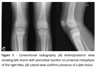
Knee radiography evidenced a lytic lesion with periosteal reaction on the proximal metaphysis of the right tibia (<a href="#f1">Figure 1</a>).</p>     ]]></body>
<body><![CDATA[<p>&nbsp;</p>    <p align="center"><a name="f1"></a><img src="/img/revistas/nas/v29n3/29n3a07f1.jpg"/></p>    
<p>&nbsp;</p>      <p>
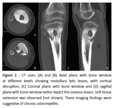
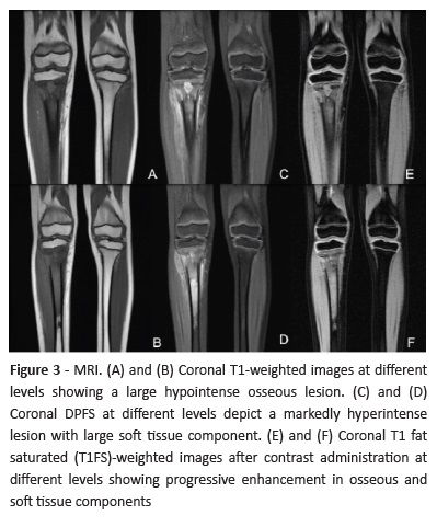
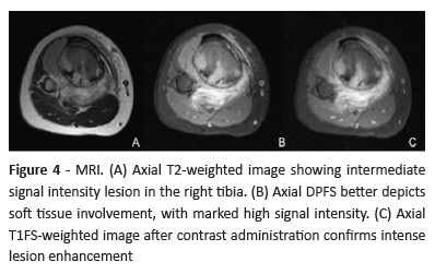
One week later, computed tomography (CT) was performed, uncovering a lytic bone lesion with soft tissue component, suggesting chronic osteomyelitis (<a href="#f2">Figure 2</a>). In the same day, magnetic resonance imaging (MRI) showed a locally aggressive lesion and findings suggestive of bone tumour (<a href="#f3">Figures 3</a> and <a href="#f4">4</a>).</p>     <p>&nbsp;</p>    <p align="center"><a name="f2"></a><img src="/img/revistas/nas/v29n3/29n3a07f2.jpg"/></p>    
<p>&nbsp;</p>    <p align="center"><a name="f3"></a><img src="/img/revistas/nas/v29n3/29n3a07f3.jpg"/></p>    
<p>&nbsp;</p>    <p align="center"><a name="f4"></a><img src="/img/revistas/nas/v29n3/29n3a07f4.jpg"/></p>    
]]></body>
<body><![CDATA[<p>&nbsp;</p>     <p>
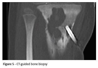
With this clinical picture, lesion biopsy was considered mandatory and CT-guided biopsy performed two weeks after admission (<a href="#f5">Figure 5</a>). Histopathological result was compatible with chronic osteomyelitis, but this finding was not consistent with imaging findings. Another biopsy was therefore performed, revealing acute leukemia. </p>     <p>&nbsp;</p>    <p align="center"><a name="f5"></a><img src="/img/revistas/nas/v29n3/29n3a07f5.jpg"/></p>    
<p>&nbsp;</p>     <p>Diagnostic workup included bone marrow aspirate, which revealed normal bone marrow morphological findings and no phenotypic evidence of acute leukemia.</p>      <p>Chemotherapy was started, with clinical response after the first cycle.</p>     <p><b>Discussion</b></p>     <p>The authors describe the clinical case of an isolated MS without evidence of leukemic involvement at diagnosis.</p>      <p>MS has been proposed to origin in bone and periosteum, as myeloid progenitors are close to the bone marrow and can thus proliferate at these sites. Cells are carried through the bone marrow to the periosteum, particularly involving the skull, orbit, sternum, spine, sacrum, and long bones. Surrounding organs can become involved by hematogenous spread from these sites.<sup>4,6</sup> MS can be found in any part of the body, either in single or multiple locations, and either concurrently or sequentially.<sup>3,4</sup> Orbit and subcutaneous tissue are the most commonly affected locations. MS may also be identified in paranasal sinuses, lymph nodes, bone, spine, brain, pleural and peritoneal cavities, breast, thyroid, salivary glands, small bowel, lung, and various pelvic organs.</p>      ]]></body>
<body><![CDATA[<p>MS is frequently asymptomatic. When symptoms are present, clinical presentation depends on the location.<sup>4,7</sup> In the present case, an isolated bone lesion on the right tibia was clinically evident as gait claudication.</p>      <p>Some clinical reports have described de novo MS without leukemic involvement at diagnosis in atypical sites &#8722; including the maxillary sinus, mandible, spinal canal, and intra-abdominally &#8722; in children aged two to nine years old.<sup>8-11</sup></p>      <p> In the present case, no progression to acute leukemia occurred during nine months. Several other clinical reports describe MS in unusual sites, in a sub-leukemic setting or with subsequent progression to acute leukemia. Bain et al reported the case of a four-month-old female diagnosed with de novo cutaneous MS, with cutaneous and bone marrow involvement one year later.<sup>7</sup> Ingale and colleagues described a case of parotid gland MS in a four-year-old male with sub-leukemic acute myeloid leukemia (AML).<sup>5</sup> Krause et al reported the case of a 16-year-old male with pericardium/heart-isolated MS relapsing in one year.<sup>12</sup></p>      <p>At CT, lesions are slightly hypodense to muscle.<sup>4</sup> After contrast administration, lesions homogeneously and more avidly enhance than muscle.<sup>3</sup> In this case, CT findings were suggestive of chronic osteomyelitis, with lytic osseous lesions and soft tissue component.</p>      <p>MRI depicts an isointense-to-bone-marrow lesion on both T1and T2-weighted images (T1WI and T2WI), with enhancement after contrast administration.<sup>4</sup> In this study, MRI raised suspicion of a tumor lesion, with intermediate signal intensity in T2WI, low signal intensity in T1WI, high signal intensity in fat suppressed proton density weighted (DPFS), and avid contrast enhancement.</p>      <p>In children, the differential diagnosis of radiographic MS includes metastatic neuroblastoma, Ewing&#8217;s sarcoma, eosinophilic granuloma, and primitive neuroectodermal tumor.<sup>4</sup></p>      <p>Several chromosomal abnormalities have been associated with myeloid sarcomas, the most common being t(8;21)(q22;q22), an abnormality shared with AML.<sup>5</sup></p>      <p>Although several therapeutic options are available (as systemic chemotherapy, surgical resection, radiotherapy, hematopoietic stem cell transplantation, or a combination of these), MS therapeutic algorithm is unclear. In isolated MS, recommendation is to start AML conventional chemotherapy as soon as possible, as untreated MS almost invariably progresses to AML.<sup>2</sup> In the present case, chemotherapy was selected, with clinical response after the first cycle.</p>      <p>Data regarding MS prognosis is controversial, with the condition being linked to poor prognosis.<sup>2,5,11</sup></p>     <p><b>Conclusion</b></p>     ]]></body>
<body><![CDATA[<p>MS is a rare entity, and isolated forms are even rarer. Isolated MS is frequently misdiagnosed because it is usually not considered in the initial differential diagnosis of osteolytic lesions. Peripheral smear and bone marrow examination are crucial to evaluate leukemic involvement.</p>     <p>&nbsp;</p>     <p><b>REFERENCES</b></p>     <!-- ref --><p>1. Swerdlow SH. Campo E, Pileri SA, Harris NL, Stein H, Siebert R, <i>et al</i>. WHO classification of tumours of haematopoietic and lymphoid tissues, fourth edition. Lyon: IARC. 2008:140-1.    &nbsp;&nbsp;&nbsp;&nbsp;&nbsp;&nbsp;&nbsp;&nbsp;[&#160;<a href="javascript:void(0);" onclick="javascript: window.open('/scielo.php?script=sci_nlinks&ref=1120420&pid=S0872-0754202000030000700001&lng=','','width=640,height=500,resizable=yes,scrollbars=1,menubar=yes,');">Links</a>&#160;]<!-- end-ref --></p>      <!-- ref --><p>2. Samborska M, Derwich K, Skalska-Sadowska J, Kurzawa P, Wachowiak J. Myeloid sarcoma in children - diagnostic and therapeutic difficulties. Contem Oncol . 2016; 20:444-8.    &nbsp;&nbsp;&nbsp;&nbsp;&nbsp;&nbsp;&nbsp;&nbsp;[&#160;<a href="javascript:void(0);" onclick="javascript: window.open('/scielo.php?script=sci_nlinks&ref=1120422&pid=S0872-0754202000030000700002&lng=','','width=640,height=500,resizable=yes,scrollbars=1,menubar=yes,');">Links</a>&#160;]<!-- end-ref --></p>      <!-- ref --><p>3. Pui MH, Fletcher BD, Langston JW. Granulocytic Sarcoma in Childhood Leukemia: Imaging Features. Radiology. 1994; 190:698-702.    &nbsp;&nbsp;&nbsp;&nbsp;&nbsp;&nbsp;&nbsp;&nbsp;[&#160;<a href="javascript:void(0);" onclick="javascript: window.open('/scielo.php?script=sci_nlinks&ref=1120424&pid=S0872-0754202000030000700003&lng=','','width=640,height=500,resizable=yes,scrollbars=1,menubar=yes,');">Links</a>&#160;]<!-- end-ref --></p>      <p>4. Guermazi A, Feger C, Rousselot P, Merad M, Benchaib N, Bourrier P. Granulocytic Sarcoma (Chloroma): Imaging Findings in Adults and Children. AJR. 2002; 178:319-25.</p>      ]]></body>
<body><![CDATA[<!-- ref --><p>5. Ingale Y, Patil T, Chaudhari P, Routray S, Agrawal M. Granulocytic Sarcoma of Parotid Gland in a 4-Year-Old Child with Subleukemic AML: A Diagnostic Challenge! Case Rep Otolaryngol. 2013; 2013:321289.    &nbsp;&nbsp;&nbsp;&nbsp;&nbsp;&nbsp;&nbsp;&nbsp;[&#160;<a href="javascript:void(0);" onclick="javascript: window.open('/scielo.php?script=sci_nlinks&ref=1120427&pid=S0872-0754202000030000700005&lng=','','width=640,height=500,resizable=yes,scrollbars=1,menubar=yes,');">Links</a>&#160;]<!-- end-ref --></p>      <!-- ref --><p>6. Gupta AJ, Mandal S, Gupta R, Khurana N, Gulati A. Myeloid Sarcoma Presenting as Nasal and Orbital Mass: An Initial Manifestation od an Acute Myeloid Leukaemia. J Clin Diagn Res. 2017; 11:ED24-6.    &nbsp;&nbsp;&nbsp;&nbsp;&nbsp;&nbsp;&nbsp;&nbsp;[&#160;<a href="javascript:void(0);" onclick="javascript: window.open('/scielo.php?script=sci_nlinks&ref=1120429&pid=S0872-0754202000030000700006&lng=','','width=640,height=500,resizable=yes,scrollbars=1,menubar=yes,');">Links</a>&#160;]<!-- end-ref --></p>      <!-- ref --><p>7. Bain EE, Rothman I, Lin L. De novo myeloid sarcoma in a 4-month-old infant: a case report and review of the literature. J Cutan Pathol . 2013; 40:321-25.    &nbsp;&nbsp;&nbsp;&nbsp;&nbsp;&nbsp;&nbsp;&nbsp;[&#160;<a href="javascript:void(0);" onclick="javascript: window.open('/scielo.php?script=sci_nlinks&ref=1120431&pid=S0872-0754202000030000700007&lng=','','width=640,height=500,resizable=yes,scrollbars=1,menubar=yes,');">Links</a>&#160;]<!-- end-ref --> </p>      <!-- ref --><p>8. Sharma A, Singh HP, Gupta AA, Garg P, Moon NJ, Chavan R. Granulocytic sarcoma in non-leukaemic child involving maxillary sinus with long term follow up: A rare case report. Ann Maxillofac Surg. 2014; 4: 90-5.    &nbsp;&nbsp;&nbsp;&nbsp;&nbsp;&nbsp;&nbsp;&nbsp;[&#160;<a href="javascript:void(0);" onclick="javascript: window.open('/scielo.php?script=sci_nlinks&ref=1120433&pid=S0872-0754202000030000700008&lng=','','width=640,height=500,resizable=yes,scrollbars=1,menubar=yes,');">Links</a>&#160;]<!-- end-ref --> </p>      <!-- ref --><p>9. Sengupta M, Das I, Chatterjee U, Majumdar B. De novo myeloid sarcoma involving mandible in a child: Report of a rare occurrence. J Oral Maxillofac Pathol. 2016; 20:304-7.    &nbsp;&nbsp;&nbsp;&nbsp;&nbsp;&nbsp;&nbsp;&nbsp;[&#160;<a href="javascript:void(0);" onclick="javascript: window.open('/scielo.php?script=sci_nlinks&ref=1120435&pid=S0872-0754202000030000700009&lng=','','width=640,height=500,resizable=yes,scrollbars=1,menubar=yes,');">Links</a>&#160;]<!-- end-ref --></p>      ]]></body>
<body><![CDATA[<!-- ref --><p>10. Shiozawa Y, Kiyokawa N, Saito M, Fujimoto J, Hata JI, Yamashiro Y. Granulocytic sarcoma of the spine in a child without bone marrow involvement: a case report and literature review . Eur J Pediatr. 2005; 164:616-20.    &nbsp;&nbsp;&nbsp;&nbsp;&nbsp;&nbsp;&nbsp;&nbsp;[&#160;<a href="javascript:void(0);" onclick="javascript: window.open('/scielo.php?script=sci_nlinks&ref=1120437&pid=S0872-0754202000030000700010&lng=','','width=640,height=500,resizable=yes,scrollbars=1,menubar=yes,');">Links</a>&#160;]<!-- end-ref --></p>      <!-- ref --><p>11. Rénard C, Girard S, Pracros JP, Dijoud F, André JM, Mialou V, <i>et al</i>. La sarcome granulocytaire, un diagnostic à connaître: à propos de 3 observations pédiatriques. Archives de Pédiatrie. 2010; 17:149-53.    &nbsp;&nbsp;&nbsp;&nbsp;&nbsp;&nbsp;&nbsp;&nbsp;[&#160;<a href="javascript:void(0);" onclick="javascript: window.open('/scielo.php?script=sci_nlinks&ref=1120439&pid=S0872-0754202000030000700011&lng=','','width=640,height=500,resizable=yes,scrollbars=1,menubar=yes,');">Links</a>&#160;]<!-- end-ref --></p>      <!-- ref --><p>12. Krause, J. Granulocytic sarcoma preceding acute leukemia. Cancer . 1979; 44:1017-21.    &nbsp;&nbsp;&nbsp;&nbsp;&nbsp;&nbsp;&nbsp;&nbsp;[&#160;<a href="javascript:void(0);" onclick="javascript: window.open('/scielo.php?script=sci_nlinks&ref=1120441&pid=S0872-0754202000030000700012&lng=','','width=640,height=500,resizable=yes,scrollbars=1,menubar=yes,');">Links</a>&#160;]<!-- end-ref --></p>     <p>&nbsp;</p>     <p><a href="#topc0">Endere&ccedil;o para correspond&ecirc;ncia</a> | <a href="#topc0">Direcci&oacute;n para correspondencia</a> | <a href="#topc0">Correspondence</a><a name="c0"></a></p>     <p>Maria Ana Serrado    <br> Department of Radiology    ]]></body>
<body><![CDATA[<br> Hospital Central do Funchal     <br> Av. Luís de Camões 6180,    <br> 9000-177 Funchal, Portugal    <br> Email: <a href="mailto:m_serrado@hotmail.com">m_serrado@hotmail.com</a></p>     <p>&nbsp;</p>     <p>Received for publication: 15.01.2019. Accepted in revised form: 10.12.2019</p>      ]]></body><back>
<ref-list>
<ref id="B1">
<label>1</label><nlm-citation citation-type="book">
<person-group person-group-type="author">
<name>
<surname><![CDATA[Swerdlow]]></surname>
<given-names><![CDATA[SH]]></given-names>
</name>
<name>
<surname><![CDATA[Campo]]></surname>
<given-names><![CDATA[E]]></given-names>
</name>
<name>
<surname><![CDATA[Pileri]]></surname>
<given-names><![CDATA[SA]]></given-names>
</name>
<name>
<surname><![CDATA[Harris]]></surname>
<given-names><![CDATA[NL]]></given-names>
</name>
<name>
<surname><![CDATA[Stein]]></surname>
<given-names><![CDATA[H]]></given-names>
</name>
<name>
<surname><![CDATA[Siebert]]></surname>
<given-names><![CDATA[R]]></given-names>
</name>
</person-group>
<source><![CDATA[WHO classification of tumours of haematopoietic and lymphoid tissues]]></source>
<year>2008</year>
<edition>fourth</edition>
<page-range>140-1</page-range><publisher-loc><![CDATA[Lyon ]]></publisher-loc>
<publisher-name><![CDATA[IARC]]></publisher-name>
</nlm-citation>
</ref>
<ref id="B2">
<label>2</label><nlm-citation citation-type="journal">
<person-group person-group-type="author">
<name>
<surname><![CDATA[Samborska]]></surname>
<given-names><![CDATA[M]]></given-names>
</name>
<name>
<surname><![CDATA[Derwich]]></surname>
<given-names><![CDATA[K]]></given-names>
</name>
<name>
<surname><![CDATA[Skalska-Sadowska]]></surname>
<given-names><![CDATA[J]]></given-names>
</name>
<name>
<surname><![CDATA[Kurzawa]]></surname>
<given-names><![CDATA[P]]></given-names>
</name>
<name>
<surname><![CDATA[Wachowiak]]></surname>
<given-names><![CDATA[J]]></given-names>
</name>
</person-group>
<article-title xml:lang="en"><![CDATA[Myeloid sarcoma in children: diagnostic and therapeutic difficulties]]></article-title>
<source><![CDATA[Contem Oncol]]></source>
<year>2016</year>
<volume>20</volume>
<page-range>444-8</page-range></nlm-citation>
</ref>
<ref id="B3">
<label>3</label><nlm-citation citation-type="journal">
<person-group person-group-type="author">
<name>
<surname><![CDATA[Pui]]></surname>
<given-names><![CDATA[MH]]></given-names>
</name>
<name>
<surname><![CDATA[Fletcher]]></surname>
<given-names><![CDATA[BD]]></given-names>
</name>
<name>
<surname><![CDATA[Langston]]></surname>
<given-names><![CDATA[JW]]></given-names>
</name>
</person-group>
<article-title xml:lang="en"><![CDATA[Granulocytic Sarcoma in Childhood Leukemia: Imaging Features]]></article-title>
<source><![CDATA[Radiology]]></source>
<year>1994</year>
<volume>190</volume>
<page-range>698-702</page-range></nlm-citation>
</ref>
<ref id="B4">
<label>4</label><nlm-citation citation-type="journal">
<person-group person-group-type="author">
<name>
<surname><![CDATA[Guermazi]]></surname>
<given-names><![CDATA[A]]></given-names>
</name>
<name>
<surname><![CDATA[Feger]]></surname>
<given-names><![CDATA[CP]]></given-names>
</name>
<name>
<surname><![CDATA[Merad]]></surname>
<given-names><![CDATA[M]]></given-names>
</name>
<name>
<surname><![CDATA[Benchaib]]></surname>
<given-names><![CDATA[N]]></given-names>
</name>
<name>
<surname><![CDATA[Bourrier]]></surname>
<given-names><![CDATA[P]]></given-names>
</name>
</person-group>
<article-title xml:lang="en"><![CDATA[Granulocytic Sarcoma (Chloroma): Imaging Findings in Adults and Children]]></article-title>
<source><![CDATA[AJR]]></source>
<year>2002</year>
<volume>178</volume>
<page-range>319-25</page-range></nlm-citation>
</ref>
<ref id="B5">
<label>5</label><nlm-citation citation-type="journal">
<person-group person-group-type="author">
<name>
<surname><![CDATA[Ingale]]></surname>
<given-names><![CDATA[Y]]></given-names>
</name>
<name>
<surname><![CDATA[Patil]]></surname>
<given-names><![CDATA[T]]></given-names>
</name>
<name>
<surname><![CDATA[Chaudhari]]></surname>
<given-names><![CDATA[P]]></given-names>
</name>
<name>
<surname><![CDATA[Routray]]></surname>
<given-names><![CDATA[S]]></given-names>
</name>
<name>
<surname><![CDATA[Agrawal]]></surname>
<given-names><![CDATA[M]]></given-names>
</name>
</person-group>
<article-title xml:lang="en"><![CDATA[Granulocytic Sarcoma of Parotid Gland in a 4-Year-Old Child with Subleukemic AML: A Diagnostic Challenge!]]></article-title>
<source><![CDATA[Case Rep Otolaryngol]]></source>
<year>2013</year>
<volume>2013</volume>
<page-range>321289</page-range></nlm-citation>
</ref>
<ref id="B6">
<label>6</label><nlm-citation citation-type="journal">
<person-group person-group-type="author">
<name>
<surname><![CDATA[Gupta]]></surname>
<given-names><![CDATA[AJ]]></given-names>
</name>
<name>
<surname><![CDATA[Mandal]]></surname>
<given-names><![CDATA[S]]></given-names>
</name>
<name>
<surname><![CDATA[Gupta]]></surname>
<given-names><![CDATA[R]]></given-names>
</name>
<name>
<surname><![CDATA[Khurana]]></surname>
<given-names><![CDATA[N]]></given-names>
</name>
<name>
<surname><![CDATA[Gulati]]></surname>
<given-names><![CDATA[A]]></given-names>
</name>
</person-group>
<article-title xml:lang="en"><![CDATA[Myeloid Sarcoma Presenting as Nasal and Orbital Mass: An Initial Manifestation od an Acute Myeloid Leukaemia]]></article-title>
<source><![CDATA[J Clin Diagn Res]]></source>
<year>2017</year>
<volume>11</volume>
<page-range>ED24-6</page-range></nlm-citation>
</ref>
<ref id="B7">
<label>7</label><nlm-citation citation-type="journal">
<person-group person-group-type="author">
<name>
<surname><![CDATA[Bain]]></surname>
<given-names><![CDATA[EE]]></given-names>
</name>
<name>
<surname><![CDATA[Rothman]]></surname>
<given-names><![CDATA[I]]></given-names>
</name>
<name>
<surname><![CDATA[Lin]]></surname>
<given-names><![CDATA[L]]></given-names>
</name>
</person-group>
<article-title xml:lang="en"><![CDATA[De novo myeloid sarcoma in a 4-month-old infant: a case report and review of the literature]]></article-title>
<source><![CDATA[J Cutan Pathol]]></source>
<year>2013</year>
<volume>40</volume>
<page-range>321-25</page-range></nlm-citation>
</ref>
<ref id="B8">
<label>8</label><nlm-citation citation-type="journal">
<person-group person-group-type="author">
<name>
<surname><![CDATA[Sharma]]></surname>
<given-names><![CDATA[A]]></given-names>
</name>
<name>
<surname><![CDATA[Singh]]></surname>
<given-names><![CDATA[HP]]></given-names>
</name>
<name>
<surname><![CDATA[Gupta]]></surname>
<given-names><![CDATA[AA]]></given-names>
</name>
<name>
<surname><![CDATA[Garg]]></surname>
<given-names><![CDATA[P]]></given-names>
</name>
<name>
<surname><![CDATA[Moon]]></surname>
<given-names><![CDATA[NJ]]></given-names>
</name>
<name>
<surname><![CDATA[Chavan]]></surname>
<given-names><![CDATA[R]]></given-names>
</name>
</person-group>
<article-title xml:lang="en"><![CDATA[Granulocytic sarcoma in non-leukaemic child involving maxillary sinus with long term follow up: A rare case report]]></article-title>
<source><![CDATA[Ann Maxillofac Surg]]></source>
<year>2014</year>
<volume>4</volume>
<page-range>90-5</page-range></nlm-citation>
</ref>
<ref id="B9">
<label>9</label><nlm-citation citation-type="journal">
<person-group person-group-type="author">
<name>
<surname><![CDATA[Sengupta]]></surname>
<given-names><![CDATA[M]]></given-names>
</name>
<name>
<surname><![CDATA[Das]]></surname>
<given-names><![CDATA[I]]></given-names>
</name>
<name>
<surname><![CDATA[Chatterjee]]></surname>
<given-names><![CDATA[U]]></given-names>
</name>
<name>
<surname><![CDATA[Majumdar]]></surname>
<given-names><![CDATA[B]]></given-names>
</name>
</person-group>
<article-title xml:lang="en"><![CDATA[De novo myeloid sarcoma involving mandible in a child: Report of a rare occurrence]]></article-title>
<source><![CDATA[J Oral Maxillofac Pathol]]></source>
<year>2016</year>
<volume>20</volume>
<page-range>304-7</page-range></nlm-citation>
</ref>
<ref id="B10">
<label>10</label><nlm-citation citation-type="journal">
<person-group person-group-type="author">
<name>
<surname><![CDATA[Shiozawa]]></surname>
<given-names><![CDATA[Y]]></given-names>
</name>
<name>
<surname><![CDATA[Kiyokawa]]></surname>
<given-names><![CDATA[N]]></given-names>
</name>
<name>
<surname><![CDATA[Saito]]></surname>
<given-names><![CDATA[M]]></given-names>
</name>
<name>
<surname><![CDATA[Fujimoto]]></surname>
<given-names><![CDATA[J]]></given-names>
</name>
<name>
<surname><![CDATA[Hata]]></surname>
<given-names><![CDATA[JI]]></given-names>
</name>
<name>
<surname><![CDATA[Yamashiro]]></surname>
<given-names><![CDATA[Y]]></given-names>
</name>
</person-group>
<article-title xml:lang="en"><![CDATA[Granulocytic sarcoma of the spine in a child without bone marrow involvement: a case report and literature review]]></article-title>
<source><![CDATA[Eur J Pediatr]]></source>
<year>2005</year>
<volume>164</volume>
<page-range>616-20</page-range></nlm-citation>
</ref>
<ref id="B11">
<label>11</label><nlm-citation citation-type="journal">
<person-group person-group-type="author">
<name>
<surname><![CDATA[Rénard]]></surname>
<given-names><![CDATA[C]]></given-names>
</name>
<name>
<surname><![CDATA[Girard]]></surname>
<given-names><![CDATA[S]]></given-names>
</name>
<name>
<surname><![CDATA[Pracros]]></surname>
<given-names><![CDATA[JP]]></given-names>
</name>
<name>
<surname><![CDATA[Dijoud]]></surname>
<given-names><![CDATA[F]]></given-names>
</name>
<name>
<surname><![CDATA[André]]></surname>
<given-names><![CDATA[JM]]></given-names>
</name>
<name>
<surname><![CDATA[Mialou]]></surname>
<given-names><![CDATA[V]]></given-names>
</name>
</person-group>
<article-title xml:lang="en"><![CDATA[La sarcome granulocytaire, un diagnostic à connaître: à propos de 3 observations pédiatriques]]></article-title>
<source><![CDATA[Archives de Pédiatrie]]></source>
<year>2010</year>
<volume>17</volume>
<page-range>149-53</page-range></nlm-citation>
</ref>
<ref id="B12">
<label>12</label><nlm-citation citation-type="journal">
<person-group person-group-type="author">
<name>
<surname><![CDATA[Krause]]></surname>
<given-names><![CDATA[J]]></given-names>
</name>
</person-group>
<article-title xml:lang="en"><![CDATA[Granulocytic sarcoma preceding acute leukemia]]></article-title>
<source><![CDATA[Cancer]]></source>
<year>1979</year>
<volume>44</volume>
<page-range>1017-21</page-range></nlm-citation>
</ref>
</ref-list>
</back>
</article>
