<?xml version="1.0" encoding="ISO-8859-1"?><article xmlns:mml="http://www.w3.org/1998/Math/MathML" xmlns:xlink="http://www.w3.org/1999/xlink" xmlns:xsi="http://www.w3.org/2001/XMLSchema-instance">
<front>
<journal-meta>
<journal-id>0872-671X</journal-id>
<journal-title><![CDATA[Medicina Interna]]></journal-title>
<abbrev-journal-title><![CDATA[Medicina Interna]]></abbrev-journal-title>
<issn>0872-671X</issn>
<publisher>
<publisher-name><![CDATA[Sociedade Portuguesa de Medicina Interna]]></publisher-name>
</publisher>
</journal-meta>
<article-meta>
<article-id>S0872-671X2017000100013</article-id>
<title-group>
<article-title xml:lang="pt"><![CDATA[O Que Esconde a Pele?]]></article-title>
<article-title xml:lang="en"><![CDATA[What Does the Skin Hide?]]></article-title>
</title-group>
<contrib-group>
<contrib contrib-type="author">
<name>
<surname><![CDATA[Campos]]></surname>
<given-names><![CDATA[Sara]]></given-names>
</name>
<xref ref-type="aff" rid="A1"/>
</contrib>
<contrib contrib-type="author">
<name>
<surname><![CDATA[João]]></surname>
<given-names><![CDATA[Alexandre]]></given-names>
</name>
<xref ref-type="aff" rid="A1"/>
</contrib>
</contrib-group>
<aff id="AA1">
<institution><![CDATA[,Centro Hospitalar Lisboa Central Hospital dos Capuchos Serviço Dermatovenereologia]]></institution>
<addr-line><![CDATA[Lisboa ]]></addr-line>
<country>Portugal</country>
</aff>
<pub-date pub-type="pub">
<day>00</day>
<month>03</month>
<year>2017</year>
</pub-date>
<pub-date pub-type="epub">
<day>00</day>
<month>03</month>
<year>2017</year>
</pub-date>
<volume>24</volume>
<numero>1</numero>
<fpage>43</fpage>
<lpage>43</lpage>
<copyright-statement/>
<copyright-year/>
<self-uri xlink:href="http://scielo.pt/scielo.php?script=sci_arttext&amp;pid=S0872-671X2017000100013&amp;lng=en&amp;nrm=iso"></self-uri><self-uri xlink:href="http://scielo.pt/scielo.php?script=sci_abstract&amp;pid=S0872-671X2017000100013&amp;lng=en&amp;nrm=iso"></self-uri><self-uri xlink:href="http://scielo.pt/scielo.php?script=sci_pdf&amp;pid=S0872-671X2017000100013&amp;lng=en&amp;nrm=iso"></self-uri><kwd-group>
<kwd lng="pt"><![CDATA[Erupções Liquenóides]]></kwd>
<kwd lng="pt"><![CDATA[Tuberculose Cutânea]]></kwd>
<kwd lng="en"><![CDATA[Lichenoid Eruptions]]></kwd>
<kwd lng="en"><![CDATA[Tuberculosis, Cutaneous]]></kwd>
</kwd-group>
</article-meta>
</front><body><![CDATA[ <p align="right"><b>IMAGENS EM MEDICINA</b> / IMAGES IN MEDICINE</p>       <p><b>O Que Esconde a Pele?</b></p>       <p><b>What Does the Skin Hide?</b></p>       <p><b>Sara Campos, Alexandre João</b></p>     <p>Serviço Dermatovenereologia, Hospital dos Capuchos - Centro Hospitalar Lisboa Central, Lisboa, Portugal</p>     <p><a name="topc0"></a><a href="#c0">Correspondência</a> </p>     <p>&nbsp;</p>      <p><b>Palavras-chave:</b>Erupções Liquenóides; Tuberculose Cutânea.</p>      <p>&nbsp;</p> <hr/>     <p><b>Keywords:</b>Lichenoid Eruptions; Tuberculosis, Cutaneous</p>     ]]></body>
<body><![CDATA[<p>&nbsp;</p> <hr/>      <p>&nbsp;</p>     <p>
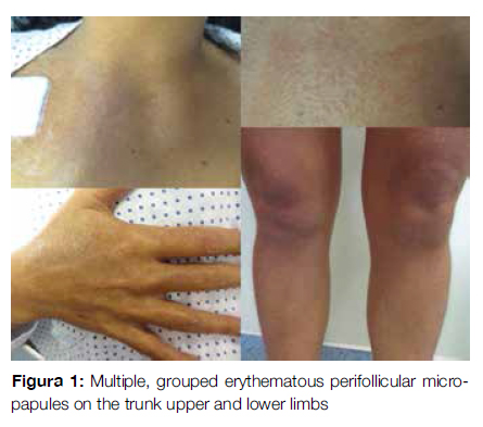
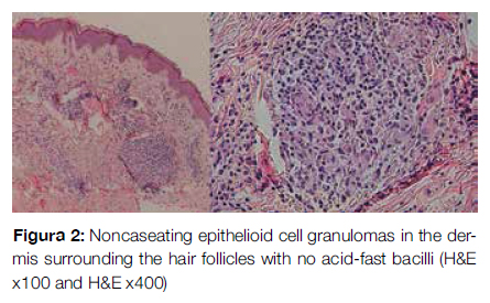
A 44-year-old woman under investigation due to a chronic granulomatous uveitis presented in our department with a one-month history of a non-pruritic skin rash. Cutaneous examination revealed multiple, grouped erythematous perifollicular micropapules over the trunk, limbs and face (<a href="#f1">Fig. 1</a>). No other symptoms were present and the remaining physical examination was normal. The skin biopsy showed non-caseating epithelioid cell granulomas in the dermis surrounding the hair follicles with no acid-fast bacilli (<a href="#f2">Fig. 2</a>). Taking together the history of chronic granulomatous uveitis and the skin biopsy result, tuberculosis infection study was performed. A positive interferon-gamma release assay and tuberculin skin test were present. Analytic study, thorax x-ray and axial tomography scan were normal. The diagnosis of lichen scrofulosorum and tubercular uveitis was established. The patient was treated with antituberculous therapy with complete resolution of the skin rash after 1 month.</p>     <p>&nbsp;</p> <a name="f1"> <img src="/img/revistas/mint/v24n1/24n1a13f1.jpg">     
<p>&nbsp;</p>     <p>&nbsp;</p> <a name="f2"> <img src="/img/revistas/mint/v24n1/24n1a13f2.jpg">     
<p>&nbsp;</p>     <p>Tuberculosis (TB) is an important cause of morbidity and mortality worldwide and usually affects the lungs, but can also affect other organs.<sup>1</sup> Tuberculous uveitis is a rare cause of extra-pulmonary TB and usually presents as chronic granulomatous uveitis. Because of the difficulty in obtaining microbiologic evidence, in nearly all reported cases, the diagnosis of intraocular TB is only presumptive.<sup>2</sup> Tuberculids develops as a hypersensitive immunologic reaction in the skin due to an occult internal focus of tuberculosis. These eruptive lesions are due to hematogenous dissemination of bacilli in a host with a high degree of immunity against Mycobacterium tuberculosis.<sup>3</sup> Lichen scrofulosorum (LS) is a rare tuberculid and can mimic several dermatologic diseases, so a high index of suspicion is required for the diagnosis.<sup>4,5</sup> This patient had two rare diseases simultaneously: intraocular TB and LS. The authors present this case to highlights LS as an important marker of undetected tuberculosis.</p>     <p>&nbsp;</p>     <p><b>Referências</b></p>     ]]></body>
<body><![CDATA[<p>1. Yates VM. Mycobacterial infections. In: Burns T, Breatnach S, Cox N, Griffiths C, editors. Rook´s Textbook of Dermatology. 8th ed. Oxford: Blackwell Science; 2010. p. 31.21–2.</p>     <!-- ref --><p>2. Gupta V, Gupta A, Rao NA. Intraocular tuberculosis: An update. Surv Ophthalmol. 2007;52:561–87.    &nbsp;&nbsp;&nbsp;&nbsp;&nbsp;&nbsp;&nbsp;&nbsp;[&#160;<a href="javascript:void(0);" onclick="javascript: window.open('/scielo.php?script=sci_nlinks&ref=1949455&pid=S0872-671X201700010001300002&lng=','','width=640,height=500,resizable=yes,scrollbars=1,menubar=yes,');">Links</a>&#160;]<!-- end-ref --></p>     <!-- ref --><p>3. Hallensleben ND, Vries HJ, Lettinga KD, Scherpbier HJ. Tuberculids: cutaneous indicator diseases of Mycobacterium tuberculosis infection in young patients. J Eur Acad Dermatol Venereol. 2016;30:1590-3.    &nbsp;&nbsp;&nbsp;&nbsp;&nbsp;&nbsp;&nbsp;&nbsp;[&#160;<a href="javascript:void(0);" onclick="javascript: window.open('/scielo.php?script=sci_nlinks&ref=1949457&pid=S0872-671X201700010001300003&lng=','','width=640,height=500,resizable=yes,scrollbars=1,menubar=yes,');">Links</a>&#160;]<!-- end-ref --></p>     <!-- ref --><p>4. Singal A, Pandhi D. Lichen scrofulosorum and endometrial tuberculosis: a novel association. Int J Dermatol. 2016;55:322-4.    &nbsp;&nbsp;&nbsp;&nbsp;&nbsp;&nbsp;&nbsp;&nbsp;[&#160;<a href="javascript:void(0);" onclick="javascript: window.open('/scielo.php?script=sci_nlinks&ref=1949459&pid=S0872-671X201700010001300004&lng=','','width=640,height=500,resizable=yes,scrollbars=1,menubar=yes,');">Links</a>&#160;]<!-- end-ref --></p>     <!-- ref --><p>5. Thami GP, Kaur S, Kanwar AJ, Mohan H. Lichen scrofulosorum: A rare manifestation of a common disease. Pediatr Dermatol. 2002;9:22–6.    &nbsp;&nbsp;&nbsp;&nbsp;&nbsp;&nbsp;&nbsp;&nbsp;[&#160;<a href="javascript:void(0);" onclick="javascript: window.open('/scielo.php?script=sci_nlinks&ref=1949461&pid=S0872-671X201700010001300005&lng=','','width=640,height=500,resizable=yes,scrollbars=1,menubar=yes,');">Links</a>&#160;]<!-- end-ref --></p>      <p>&nbsp;</p>     ]]></body>
<body><![CDATA[<p><a name="c0"></a><a href="#topc0">Correspondência</a>: Sara Campos <a href="mailto:saraazevedocampos@gmail.com">saraazevedocampos@gmail.com</a>     <br> Serviço Dermatovenereologia, Hospital dos Capuchos - Centro Hospitalar Lisboa Central, Lisboa, Portugal<br /> Alameda de Santo António dos Capuchos, 1169-050 Lisboa</p>     <p>&nbsp;</p>      <p>Protecção de Seres Humanos e Animais: Os autores declaram que não foram realizadas experiências em seres humanos ou animais</p>     <p>Direito à Privacidade e Consentimento Informado: Os autores declaram que nenhum dado que permita a identificação do doente aparece neste artigo.</p>     <p>Conflitos de Interesse: Os autores declaram a inexistência de conflitos de interesse na realização do presente trabalho</p>     <p>Fontes de Financiamento: Não existiram fontes externas de financiamento para a realização deste artigo</p>     <p>&nbsp;</p>     <p>Recebido: 31/10/2016</p>     <p>Aceite:  08/11/2016</p>     ]]></body>
<body><![CDATA[ ]]></body><back>
<ref-list>
<ref id="B1">
<label>1</label><nlm-citation citation-type="journal">
<person-group person-group-type="author">
<name>
<surname><![CDATA[Yates]]></surname>
<given-names><![CDATA[VM]]></given-names>
</name>
</person-group>
<article-title xml:lang="en"><![CDATA[Mycobacterial infections: In: Burns T, Breatnach S, Cox N, Griffiths C, editors. Rook´s Textbook of Dermatology. 8th ed]]></article-title>
<source><![CDATA[Oxford: Blackwell Science;]]></source>
<year>2010</year>
<volume>31</volume>
<page-range>21-2</page-range></nlm-citation>
</ref>
<ref id="B2">
<label>2</label><nlm-citation citation-type="journal">
<person-group person-group-type="author">
<name>
<surname><![CDATA[Gupta]]></surname>
<given-names><![CDATA[V]]></given-names>
</name>
<name>
<surname><![CDATA[Gupta]]></surname>
<given-names><![CDATA[A]]></given-names>
</name>
<name>
<surname><![CDATA[Rao]]></surname>
<given-names><![CDATA[NA]]></given-names>
</name>
</person-group>
<article-title xml:lang="en"><![CDATA[Intraocular tuberculosis: An update]]></article-title>
<source><![CDATA[Surv Ophthalmol]]></source>
<year>2007</year>
<volume>52</volume>
<page-range>561-87</page-range></nlm-citation>
</ref>
<ref id="B3">
<label>3</label><nlm-citation citation-type="journal">
<person-group person-group-type="author">
<name>
<surname><![CDATA[Hallensleben]]></surname>
<given-names><![CDATA[ND]]></given-names>
</name>
<name>
<surname><![CDATA[Vries]]></surname>
<given-names><![CDATA[HJ]]></given-names>
</name>
<name>
<surname><![CDATA[Lettinga]]></surname>
<given-names><![CDATA[KD]]></given-names>
</name>
<name>
<surname><![CDATA[Scherpbier]]></surname>
<given-names><![CDATA[HJ]]></given-names>
</name>
</person-group>
<article-title xml:lang="en"><![CDATA[Tuberculids: cutaneous indicator diseases of Mycobacterium tuberculosis infection in young patients]]></article-title>
<source><![CDATA[J Eur Acad Dermatol Venereol]]></source>
<year>2016</year>
<volume>30</volume>
<page-range>1590-3</page-range></nlm-citation>
</ref>
<ref id="B4">
<label>4</label><nlm-citation citation-type="journal">
<person-group person-group-type="author">
<name>
<surname><![CDATA[Singal]]></surname>
<given-names><![CDATA[A]]></given-names>
</name>
<name>
<surname><![CDATA[Pandhi]]></surname>
<given-names><![CDATA[D]]></given-names>
</name>
</person-group>
<article-title xml:lang="en"><![CDATA[Lichen scrofulosorum and endometrial tuberculosis: a novel association]]></article-title>
<source><![CDATA[Int J Dermatol]]></source>
<year>2016</year>
<volume>55</volume>
<page-range>322-4</page-range></nlm-citation>
</ref>
<ref id="B5">
<label>5</label><nlm-citation citation-type="journal">
<person-group person-group-type="author">
<name>
<surname><![CDATA[Thami]]></surname>
<given-names><![CDATA[GP]]></given-names>
</name>
<name>
<surname><![CDATA[Kaur]]></surname>
<given-names><![CDATA[S]]></given-names>
</name>
<name>
<surname><![CDATA[Kanwar]]></surname>
<given-names><![CDATA[AJ]]></given-names>
</name>
<name>
<surname><![CDATA[Mohan]]></surname>
<given-names><![CDATA[H]]></given-names>
</name>
</person-group>
<article-title xml:lang="en"><![CDATA[Lichen scrofulosorum: A rare manifestation of a common disease]]></article-title>
<source><![CDATA[Pediatr Dermatol]]></source>
<year>2002</year>
<volume>9</volume>
<page-range>22-6</page-range></nlm-citation>
</ref>
</ref-list>
</back>
</article>
