<?xml version="1.0" encoding="ISO-8859-1"?><article xmlns:mml="http://www.w3.org/1998/Math/MathML" xmlns:xlink="http://www.w3.org/1999/xlink" xmlns:xsi="http://www.w3.org/2001/XMLSchema-instance">
<front>
<journal-meta>
<journal-id>1646-107X</journal-id>
<journal-title><![CDATA[Motricidade]]></journal-title>
<abbrev-journal-title><![CDATA[Motri.]]></abbrev-journal-title>
<issn>1646-107X</issn>
<publisher>
<publisher-name><![CDATA[Edições Desafio Singular]]></publisher-name>
</publisher>
</journal-meta>
<article-meta>
<article-id>S1646-107X2015000400012</article-id>
<article-id pub-id-type="doi">10.6063/motricidade.5999</article-id>
<title-group>
<article-title xml:lang="en"><![CDATA[Influence of Subcutaneous Fat Layer in Skin Temperature]]></article-title>
<article-title xml:lang="pt"><![CDATA[Influência da Camada de Gordura Subcutânea na Temperatura da Pele]]></article-title>
</title-group>
<contrib-group>
<contrib contrib-type="author">
<name>
<surname><![CDATA[Neves]]></surname>
<given-names><![CDATA[Eduardo Borba]]></given-names>
</name>
<xref ref-type="aff" rid="A01"/>
<xref ref-type="aff" rid="A03"/>
</contrib>
<contrib contrib-type="author">
<name>
<surname><![CDATA[Vilaça-Alves]]></surname>
<given-names><![CDATA[José]]></given-names>
</name>
<xref ref-type="aff" rid="A02"/>
<xref ref-type="aff" rid="A03"/>
</contrib>
<contrib contrib-type="author">
<name>
<surname><![CDATA[Nogueira]]></surname>
<given-names><![CDATA[Isabel Regina Amorim]]></given-names>
</name>
<xref ref-type="aff" rid="A03"/>
</contrib>
<contrib contrib-type="author">
<name>
<surname><![CDATA[Reis]]></surname>
<given-names><![CDATA[Victor Machado]]></given-names>
</name>
<xref ref-type="aff" rid="A02"/>
<xref ref-type="aff" rid="A03"/>
</contrib>
</contrib-group>
<aff id="A01">
<institution><![CDATA[,Brazilian Army Research Institute of Physical Fitness  ]]></institution>
<addr-line><![CDATA[Rio de Janeiro ]]></addr-line>
<country>Brazil</country>
</aff>
<aff id="A02">
<institution><![CDATA[,Research Centre in Sports Sciences, Health Sciences and Human Development  ]]></institution>
<addr-line><![CDATA[ ]]></addr-line>
<country>Portugal</country>
</aff>
<aff id="A03">
<institution><![CDATA[,University of Trás-os-Montes and Alto Douro  ]]></institution>
<addr-line><![CDATA[ ]]></addr-line>
</aff>
<pub-date pub-type="pub">
<day>01</day>
<month>12</month>
<year>2015</year>
</pub-date>
<pub-date pub-type="epub">
<day>01</day>
<month>12</month>
<year>2015</year>
</pub-date>
<volume>11</volume>
<numero>4</numero>
<fpage>120</fpage>
<lpage>126</lpage>
<copyright-statement/>
<copyright-year/>
<self-uri xlink:href="http://scielo.pt/scielo.php?script=sci_arttext&amp;pid=S1646-107X2015000400012&amp;lng=en&amp;nrm=iso"></self-uri><self-uri xlink:href="http://scielo.pt/scielo.php?script=sci_abstract&amp;pid=S1646-107X2015000400012&amp;lng=en&amp;nrm=iso"></self-uri><self-uri xlink:href="http://scielo.pt/scielo.php?script=sci_pdf&amp;pid=S1646-107X2015000400012&amp;lng=en&amp;nrm=iso"></self-uri><abstract abstract-type="short" xml:lang="en"><p><![CDATA[The aim of this study was to determine the correlation between the subcutaneous fat layer and the temperature variation (core - skin) in two body regions, measured by infrared sensors. This study involved 43 volunteers selected among undergraduate students in physical education. The thermal images and skinfold thickness were acquired from subscapular region (SB) for male and triceps region (TR) for female volunteers, of right side. In this study, subscapular skinfold thickness was inversely correlated with skin temperature (r = -0.638, p = 0.004) and directly correlated with &#916;T [core - skin] (r = 0.653, p = 0.003). However, the results for the triceps region showed no significant correlation between triceps skinfold thickness and skin temperature or &#916;T [core - skin]. When the sample was separated into two groups by skinfold thickness, with cut off at 12mm, average comparison tests for &#916;T [core - skin] shows statistically significant difference between groups for the subscapular site (Student t test for independent sample, p value = 0.023), but not for the triceps site (Wilcoxon Signed Ranks and the Mann-Whitney tests, p value = 0.268). In short, the findings suggest that subcutaneous fat layer correlates negatively with skin temperature in subscapular region.]]></p></abstract>
<abstract abstract-type="short" xml:lang="pt"><p><![CDATA[O objetivo deste estudo foi determinar a correlação entre a camada de gordura subcutânea e a variação de temperatura (core - pele) em duas regiões do corpo, medida por sensores infravermelhos. Este estudo envolveu 43 voluntários, selecionados entre os estudantes de graduação em educação física. As imagens térmicas e dobras cutâneas foram adquiridas da região subescapular (SB) para o sexo masculino e da região tricipital (TR) para as voluntárias do sexo feminino, ambas do lado direito do corpo. Neste estudo, a espessura da prega cutânea subescapular, correlacionou-se inversamente com a temperatura da pele (r = -0,638, p = 0,004) e positivamente com a variação de temperatura [central - pele] (r = 0,653, p = 0,003). No entanto, os resultados para a região tricipital não mostrou correlação significativa com a temperatura da pele ou com a variação de temperatura [central - pele]. Quando a amostra foi separada em dois grupos, pela espessura da prega cutânea, com ponto de corte em 12mm, teste de médias de comparação para a variação de temperatura [central - pele] mostrou diferença estatisticamente significativa entre os grupos para a região subescapular (teste t de Student para amostra independente, valor de p = 0,023 ), mas não para a região tricipital (Wilcoxon Signed Ranks e os testes de Mann-Whitney, p = 0,268). Em suma, os resultados sugerem que a camada subcutânea de gordura está negativamente correlacionada com a temperatura da pele na região subescapular.]]></p></abstract>
<kwd-group>
<kwd lng="en"><![CDATA[thermography]]></kwd>
<kwd lng="en"><![CDATA[subcutaneous fat layer]]></kwd>
<kwd lng="en"><![CDATA[skin temperature]]></kwd>
<kwd lng="en"><![CDATA[human thermal physiology]]></kwd>
<kwd lng="en"><![CDATA[skinfold thickness]]></kwd>
<kwd lng="pt"><![CDATA[termografia]]></kwd>
<kwd lng="pt"><![CDATA[camada de gordura subcutânea]]></kwd>
<kwd lng="pt"><![CDATA[temperatura da pele]]></kwd>
<kwd lng="pt"><![CDATA[fisiologia humana térmica]]></kwd>
<kwd lng="pt"><![CDATA[dobras cutâneas]]></kwd>
</kwd-group>
</article-meta>
</front><body><![CDATA[ <p align="right"><font size="2" face="Verdana"><b>ORIGINAL ARTICLE</b></font></p>     <p>&nbsp;</p>     <p><font size="4" face="Verdana"><b>Influence of Subcutaneous Fat Layer in   Skin Temperature</b></font></p>     <p>&nbsp;</p>     <p><font size="3" face="Verdana"><b>Influência da Camada de   Gordura Subcutânea na Temperatura da Pele</b></font></p>     <p>&nbsp;</p>     <p>&nbsp;</p>     <p><b><font size="2" face="Verdana">Eduardo Borba Neves<sup>1,3,<a href="#end">*</a></sup><a name="topo"></a>;   José Vilaça-Alves<sup>2,3</sup>; Isabel Regina Amorim   Nogueira<sup>3</sup>; Victor Machado Reis<sup>2,3</sup></font></b></p>     <p><font size="2" face="Verdana"><sup>1</sup> <i>Brazilian   Army Research Institute of Physical Fitness, Rio de Janeiro, Brazil    <br>   </i><sup>2</sup> <i>Research Centre in Sports     Sciences, Health Sciences and Human Development, CIDESD, Portugal    ]]></body>
<body><![CDATA[<br>   </i><sup>3</sup> <i>University of Tr&aacute;s-os-Montes and Alto Douro, Vila Real, Portugal</i></font></p>     <p>&nbsp;</p>     <p>&nbsp;</p> <hr noshade size="1">     <p><font size="2" face="Verdana"><b>ABSTRACT</b></font></p>     <p><font size="2" face="Verdana">The aim   of this study was to determine the correlation between the subcutaneous fat   layer and the temperature variation (core - skin) in two body regions, measured   by infrared sensors. This study involved 43 volunteers selected among   undergraduate students in physical education. The   thermal images and skinfold thickness were acquired from subscapular region   (SB) for   male and triceps region (TR) for female volunteers, of right side. In   this study, subscapular skinfold thickness was inversely correlated with skin   temperature (r = -0.638, p = 0.004) and directly correlated with &#916;T [core – skin]   (r = 0.653, p = 0.003). However, the results for the triceps   region showed no significant correlation between triceps skinfold thickness and   skin temperature or &#916;T [core – skin].   When the sample was separated into two groups by skinfold thickness, with   cut off at 12mm, average comparison tests for &#916;T [core – skin]   shows statistically significant difference between groups for the subscapular   site (Student t test for independent sample, <i>p value</i> = 0.023), but not   for the triceps site (Wilcoxon Signed Ranks and the Mann-Whitney tests, <i>p     value</i> = 0.268). In short, the findings suggest that subcutaneous fat layer correlates negatively with skin temperature in subscapular region.</font></p>     <p><font size="2" face="Verdana"><b>Keywords: </b>thermography; subcutaneous   fat layer; skin temperature; human thermal physiology; skinfold thickness.</font></p> <hr noshade size="1">     <p><font size="2" face="Verdana"><b>RESUMO</b></font></p>     <p><font size="2" face="Verdana">O   objetivo deste estudo foi determinar a correlação entre a camada de gordura   subcutânea e a variação de temperatura (core - pele) em duas regiões do corpo,   medida por sensores infravermelhos. Este estudo envolveu 43 voluntários,   selecionados entre os estudantes de graduação em educação física. As imagens   térmicas e dobras cutâneas foram adquiridas da região subescapular (SB) para o   sexo masculino e da região tricipital (TR) para as voluntárias do sexo   feminino, ambas do lado direito do corpo. Neste estudo, a espessura da prega   cutânea subescapular, correlacionou-se inversamente com a temperatura da pele   (r = -0,638, p = 0,004) e positivamente com a variação de temperatura [central   - pele] (r = 0,653, p = 0,003). No entanto, os resultados para a região   tricipital não mostrou correlação significativa com a temperatura da pele ou   com a variação de temperatura [central - pele]. Quando a amostra foi separada   em dois grupos, pela espessura da prega cutânea, com ponto de corte em 12mm,   teste de médias de comparação para a variação de temperatura [central - pele] mostrou   diferença estatisticamente significativa entre os grupos para a região   subescapular (teste t de Student para amostra independente, valor de p = 0,023   ), mas não para a região tricipital (Wilcoxon Signed Ranks e os testes de   Mann-Whitney, p = 0,268). Em suma, os resultados sugerem que a camada   subcutânea de gordura está negativamente correlacionada com a temperatura da pele na região subescapular.</font></p>     <p><font size="2" face="Verdana"><b>Palavras-chave: </b>termografia; camada de gordura subcutânea; temperatura da pele; fisiologia humana térmica; dobras cutâneas.</font></p> <hr noshade size="1">     <p>&nbsp;</p>     ]]></body>
<body><![CDATA[<p>&nbsp;</p> <font size="3" face="Verdana"><b>INTRODUCTION</b></font><font face="Verdana">     <p><font size="2">The human   body has complex mechanisms responsible for the control of body temperature   involving thermal sensors such as the hypothalamus and efferent systems. The   constant core body temperature is essential to sustain homeostasis and   maintaining the functions and composition of tissues and fluids (Jones &amp; Plassmann,   2002).   Thermography is recognized diagnostic method since the 1980s by the American   Medical Association. This method involves the detection of infrared radiation   emitted by the skin noninvasively without exposing the patient to radiation of any kind (Neves &amp; Reis, 2014).</font></p>     <p><font size="2">Heat   dissipation is influenced by many variables. Among them, the local   vascularization and subcutaneous fat layer (Bandeira, Moura, Souza,   Nohama, &amp; Neves, 2012),   that is different in male and female subjects (Jones &amp; Plassmann, 2002).</font></p>     <p><font size="2">Chudecka and Lubkowska   (2012)   found a strong correlation between maximal oxygen consumption and a decrease in   surface temperature of the upper limbs, immediately after the physical training   of a volleyball team. With these findings, the authors suggest that   thermography can be used as an additional, noninvasive method to assess the   fitness level of the athletes. They further report that the best place for such   evaluation is the front surface of the arms and forearms, because the back side   has a greater subcutaneous fat layer, which may act as an insulator of heat generated by the body.</font></p>     <p><font size="2">Other   authors (Bandeira et al., 2012;   Jones &amp; Plassmann, 2002)   have also reported the influence of subcutaneous fat layer in the assessments   thermal images. However, they not deepened the discussion of the magnitude and   direction of this influence. Furthermore, it is known that the skin temperature   depends on the core temperature (Havenith, Smith, &amp;   Fukazawa, 2008),   so it is important to consider core temperature in analysis of fat layer influence for skin temperature when measured by infrared sensors.</font></p>     <p><font size="2">Elucidation   of these relationships can contribute to the knowledge about the human thermal   physiology and can be applied immediately in the sports training planning (Neves &amp; Reis, 2014) and in development of   products such as sporting clothing (Bogerd, Psikuta, Daanen, &amp;   Rossi, 2010).   In this sense, the aim of this study was to determine the correlation between   the subcutaneous fat layer and the temperature variation (core - skin) in two body regions, measured by infrared sensors.</font></p> </font>     <p>&nbsp;</p>     <p><font size="3" face="Verdana"><b>MATERIAL AND <b>METHODS</b></b></font></p>     <p><font size="2" face="Verdana"><b>Participants</b>    </font></p>     <p><font size="2" face="Verdana">This study   involved 43 volunteers (18 males and 25 females) selected among graduate   students in physical education and the research protocol was approved by Human   Research Ethics Committee of Campos de Andrade University Center under CAAE number 28901414.3.0000.5218.</font></p>     ]]></body>
<body><![CDATA[<p><font size="2" face="Verdana">The   inclusion criterion for participation in the study was to present tympanic   temperature (regarded as core temperature) ranging between 36.0 and 36.5 in   order to keep the homogeneity of the sample (with a slight variation in core temperature).</font></p>     <p><font size="2" face="Verdana"><b>Instrumentation and Data Acquisition</b></font></p>     <p><font size="2" face="Verdana">It was used   a thermographic camera (FLIR Systems Inc. Model SC2000); a computer (with   specific software for acquisition and processing of thermographic images   ThermaCam<sup>TM</sup> Researcher Pro 2.9); and a term digital hygrometer to   monitor the temperature and humidity of the room. The thermographic camera used   has a resolution of 320 x 240 pixels, which has sensors that measures   temperatures ranging from -20° C to +120° C. This camera has a sensitivity to   detect differences of less than 0.1°C temperature and provides accuracy of ± 1°C of the absolute temperature.</font></p>     <p><font size="2" face="Verdana">The first   part of thermal images acquisition protocol was the region of interest (ROI)   delineation using tape (which reduces the emission of infrared radiation where   the ribbon is attached and allows recognizing the markers in the thermal image)   (Neves &amp; Reis, 2014). Next, the volunteers   remained for 15 minutes in an acclimatized room with temperature of 23ºC for reaching thermal balance before the images acquisition (Bandeira et al., 2014).</font></p>     <p><font size="2" face="Verdana">The thermal   images were acquired from subscapular region (SB) for male and triceps region   (TR) for female volunteers, of right side, at the site of measurement of   skinfold thickness, as the protocol described by (Neves, Ripka, Ulbricht,   &amp; Stadnik, 2013).   The skinfolds thicknesses of these sites were assessed according to Gonçalves and Mourão (2008).</font></p>     <p><font size="2" face="Verdana">It was also   used the following equipment for the anthropometric assessment: one digital   scale (Wiso W801), with capacity of 0-180 kg and grading of 100 g; one   calibrated scientific adipometer (Cescorf); and one stadiometer (WCS Woody Compact).</font></p>     <p><font size="2" face="Verdana">The skinfold   thickness used was selected because the subscapular and the triceps site have, normally,   a large subcutaneous fat layer in man (Neves et al., 2013; Ross et al.,   1994) and women (Ross et al., 1994)), respectively.</font></p>     <p><font size="2" face="Verdana">The core   temperature (Tc) was measured by tympanic access using a Braun Thermoscan   infrared ear tympanic thermometer, because as the eardrum shares the same   arterial blood supply to the hypothalamus, the tympanic temperature may be   considered as the Tc (Potter &amp; Perry, 2006). </font></p>     <p><font size="2" face="Verdana">The values of temperature variation (core - skin) were calculated according to Equation 1.</font></p>     <p><img src="/img/revistas/mot/v11n4/11n4a12e1.jpg" width="364" height="64"></p>     
]]></body>
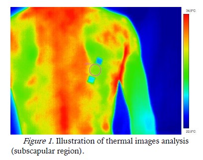
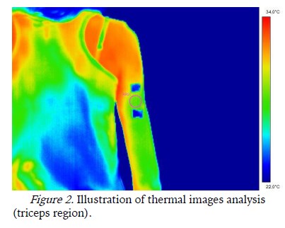
<body><![CDATA[<p><font size="2" face="Verdana">Where: &#8710;T   = temperature variation (°C), Tc = core temperature (°C), Tsk = skin temperature (°C).</font></p>     <p><font size="2" face="Verdana"><b>D</b><b>ata Processing and Analysis</b></font></p>     <p><font size="2" face="Verdana">All acquired   image were analyzed by FLIR ThermaCam<sup>TM</sup> Researcher Pro 2.9 software.   The software was set to treat the image in the colour palette midgreen (Sanches, Gamba, Souza,   Neves, &amp; Nohama, 2013)   with background temperature of 22°C and emissivity of 0.98 (Neves &amp; Reis, 2014). The circle tool was   used and the average temperature of ROI in subscapular and triceps regions was considered, as illustrated in Figures <a href="#f1">1</a> and <a href="#f2">2</a>.</font></p>     <p><a name="f1"></a></p>     <p>&nbsp;</p>     <p align="center"><img src="/img/revistas/mot/v11n4/11n4a12f1.jpg" width="408" height="321"></p>     
<p>&nbsp;</p>     <p><a name="f2"></a></p>     <p>&nbsp;</p>     <p align="center"><img src="/img/revistas/mot/v11n4/11n4a12f2.jpg" width="403" height="322"></p>     
]]></body>
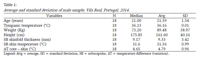
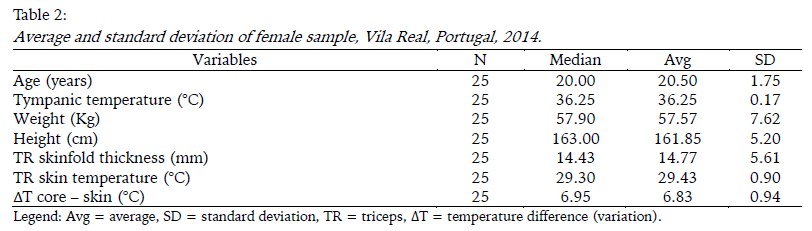
<body><![CDATA[<p>&nbsp;</p>     <p><font size="2" face="Verdana">The   Statistical analyses were performed with Statistical Package for Social   Sciences (SPSS, version 21.0). Descriptive statistics (means and SD) were used   to summarize the characteristics of the study sample, Shapiro-Wilk test was   performed to test the variable distributions, Pearson (r) and Spearman (rho)   correlation analysis was used for the main evaluations; the Student t test,   Wilcoxon Signed Ranks and the Mann-Whitney tests were applied to verify the   difference between groups. The statistical significance level was defined as p   &lt; 0.05.</font></p>     <p>&nbsp;</p> <font size="3" face="Verdana"><b>R</b><b>ESULTS</b></font><font face="Verdana">     <p><font size="2">The main characteristics of study sample was presented in Tables <a href="/img/revistas/mot/v11n4/11n4a12t1.jpg">1</a> and <a href="/img/revistas/mot/v11n4/11n4a12t2.jpg">2</a>. </font></p>     
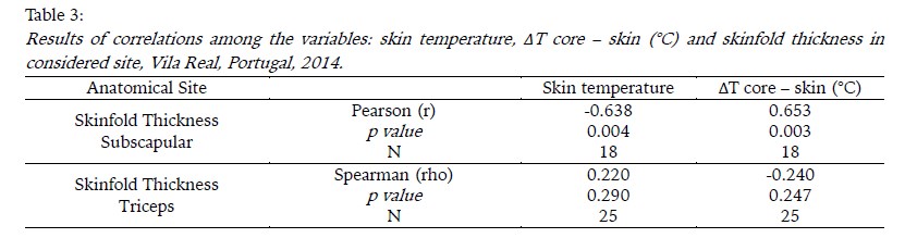
<p><font size="2">The results of   Shapiro-Wilk test showed <i>p</i> = 0.033 for &#916;T core – skin (°C) acquired at triceps. All others   variables presented p value high than 0.05. Thus, was carry out the Spearman   correlation between &#916;T core – skin (°C) and triceps skinfold thickness,   and Pearson correlation between &#916;T core – skin (°C) and triceps skinfold thickness. All results of correlations tests was showed in <a href="/img/revistas/mot/v11n4/11n4a12t3.jpg">Table 3</a>.</font></p>     
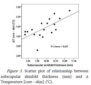
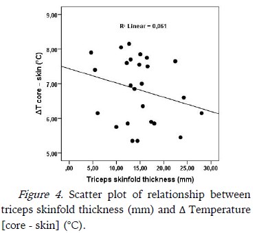
<p><font size="2">Figures <a href="#f3">3</a>  and <a href="#f4">4</a> show the scatter plot of the &#916;T   [core – skin] (°C) and skinfold thickness in each of the anatomical sites studied.</font></p>     <p><a name="f3"></a></p>     <p>&nbsp;</p>     <p align="center"><img src="/img/revistas/mot/v11n4/11n4a12f3.jpg" width="378" height="354"></p>     
<p>&nbsp;</p>     ]]></body>
<body><![CDATA[<p><a name="f4"></a></p>     <p>&nbsp;</p>     <p align="center"><img src="/img/revistas/mot/v11n4/11n4a12f4.jpg" width="377" height="340"></p>     
<p>&nbsp;</p>     <p><font size="2">When the   sample was separated into two groups by skinfold thickness, with cut off at   12mm, average comparison tests for &#916;T   [core – skin] shows   statistically significant difference between groups for the subscapular site   (Student t test for independent sample, <i>p value</i> = 0.023), but not for   the triceps site (Wilcoxon Signed Ranks and the Mann-Whitney tests, <i>p value</i> = 0.268).</font></p> </font>     <p>&nbsp;</p> <font size="3" face="Verdana"><b>DISCUSSION</b></font><font face="Verdana">     <p><font size="2">McArdle, Katch, and   Katch (2011)   and Guyton and Hall (2011) report that fat acts as   a good insulator because it has a low thermal conductivity. This study aimed to   determine the correlation between the subcutaneous fat layer and the   temperature variation (&#916;T [core – skin]) in two body regions,   measured by infrared sensors. The sample can be considered thermally   homogeneous within groups of males (subscapular thickness) and female (triceps   thickness) because shown a small standard deviation in tympanic and skin temperature. </font></p>     <p><font size="2">Although the   studied groups present gender difference, the results suggest that the   subscapular region has a higher skin temperature than triceps region. One   explanation for this may be linked to physiological maintenance processes of   life, such as the heartbeat and breathing, which provide continuous heat   generation and a large blood flow in the region subscapular (Netter, 2010; Neves   &amp; Reis, 2014; Pocock, Richards, &amp; Richards, 2013), on the other hand, in   the triceps region, at rest situation, heat is originated primarily from the blood flow, which is smaller than that of subscapular region (Netter, 2010; Pocock et al., 2013).</font></p>     <p><font size="2">In this   study, subscapular skinfold thickness was inversely correlated with skin   temperature (r = -0.638, p = 0.004) and directly correlated with &#916;T [core – skin] (r = 0.653, p = 0.003). Bandeira, Moura, Souza,   Nohama, and Neves (2012)   published similar results when reported has found significant correlation   between thigh skin temperature and thigh skinfold thickness (r = -0.488, p =   0.020) over rectus femoris muscle, in volunteers with a thigh skinfold   thickness mean of 11.40.   In the same   sense, Savastano et al. (2009) compared skin   temperature in the abdominal region into two groups (obese and normal weight)   and noted that the abdominal skin temperature of obese group was lower than in   normal-weight group, even with no difference between groups for core temperature.   The correlation (r) between abdominal skin temperature and body fat percentage   founded by Savastano et al. (2009) was 0.545. The same   authors suggest that regions of the body of greater adiposity reduce the   dissipation of central heat and the peripheral areas such as upper limbs can exercise greater heat dissipation.</font></p>     <p><font size="2">However, the   results of this study also showed no significant correlation between triceps   skinfold thickness and skin temperature or &#916;T [core – skin]. And, observing the analysis of   groups when stratified into two subgroups (larger skinfolds and smaller than   12mm), it is noticed that there is statistically difference between the   subgroups of subscapular region, but the same does not happen in the triceps   region. This fact maybe can explained because the women show biceps skinfold   thickness with coefficients of variation greater than those found in other   places of skinfold thickness assessment (Amaral et al., 2011;   Kannieappan, Deussen, Grivell, Yelland, &amp; Dodd, 2013). This can also lead to changes in places of greater heat dissipation of arm (Savastano et al., 2009). </font></p>     ]]></body>
<body><![CDATA[<p><font size="2">Regarding   the study limitations, it can be pointed the missing of measurement of muscle   mass under the ROIs studied, since the muscle mass can influence the skin   temperature (Neves,   Bandeira, Ulbricht, Vilaça-Alves, &amp; Reis, 2015). These findings just can be   generalized to men's trunk region and to women's triceps region, in subjects   with similar range of skinfold thickness. Others studies are necessary to widen the knowledge about the influence of subcutaneous fat layer in whole body. </font></p> </font>     <p>&nbsp;</p>     <p><font size="3" face="Verdana"><b>CONCLUSIONS</b></font></p>     <p><font size="2" face="Verdana">In short,   the findings suggest that subcutaneous fat layer correlates negatively with   skin temperature in subscapular region and, considering anatomical   characteristics, this result may be generalizable to other regions of the   trunk. No significant correlations between the skin temperature and   subcutaneous fat layer for triceps region were observed. These results suggest   that the variability of the subcutaneous fat layer distribution can change the   regions of heat dissipation. Future studies must to consider the inclusion of   subcutaneous fat layer as a covariable in the statistical analysis, since it influences the skin temperature acquired by infrared sensors.</font></p>     <p>&nbsp;</p>     <p><font size="3" face="Verdana"><b>REFERENCES</b> </font></p>     <!-- ref --><p><font size="2" face="Verdana">Amaral, T. F.,   Restivo, M. T., Guerra, R. S., Marques, E., Chousal, M. F., &amp; Mota, J.   (2011). Accuracy   of a digital skinfold system for measuring skinfold thickness and estimating   body fat. <i>The British Journal of Nutrition</i>, <i>105</i>(3), 478–484. <a href="http://doi.org/10.1017/S0007114510003727" target="_blank">http://doi.org/10.1017/S0007114510003727</a></font>&nbsp;&nbsp;&nbsp;&nbsp;&nbsp;&nbsp;&nbsp;&nbsp;[&#160;<a href="javascript:void(0);" onclick="javascript: window.open('/scielo.php?script=sci_nlinks&ref=351675&pid=S1646-107X201500040001200001&lng=','','width=640,height=500,resizable=yes,scrollbars=1,menubar=yes,');">Links</a>&#160;]<!-- end-ref --><!-- ref --><p><font size="2" face="Verdana">Bandeira, F., Moura, M. A. M. de, Souza,   M. A. de, Nohama, P., &amp; Neves, E. B. (2012). Can thermography aid in the   diagnosis of muscle injuries in soccer athletes? <i>Revista Brasileira     de Medicina Do Esporte</i>, <i>18</i>(4), 246–251. <a href="http://doi.org/10.1590/S1517-86922012000400006" target="_blank">http://doi.org/10.1590/S1517-86922012000400006</a></font>&nbsp;&nbsp;&nbsp;&nbsp;&nbsp;&nbsp;&nbsp;&nbsp;[&#160;<a href="javascript:void(0);" onclick="javascript: window.open('/scielo.php?script=sci_nlinks&ref=351676&pid=S1646-107X201500040001200002&lng=','','width=640,height=500,resizable=yes,scrollbars=1,menubar=yes,');">Links</a>&#160;]<!-- end-ref --><!-- ref --><p><font size="2" face="Verdana">Bandeira, F., Neves, E. B., Moura, M. A.   M. de, Nohama, P., Bandeira, F., Neves, E. B., … Nohama, P. (2014).   The thermography in support for diagnosis of muscle injury in sport. <i>Revista Brasileira     de Medicina Do Esporte</i>, <i>20</i>(1), 59–64. <a href="http://doi.org/10.1590/S1517-86922014000100012" target="_blank">http://doi.org/10.1590/S1517-86922014000100012</a></font>&nbsp;&nbsp;&nbsp;&nbsp;&nbsp;&nbsp;&nbsp;&nbsp;[&#160;<a href="javascript:void(0);" onclick="javascript: window.open('/scielo.php?script=sci_nlinks&ref=351677&pid=S1646-107X201500040001200003&lng=','','width=640,height=500,resizable=yes,scrollbars=1,menubar=yes,');">Links</a>&#160;]<!-- end-ref --><!-- ref --><p><font size="2" face="Verdana">Bogerd, N., Psikuta, A., Daanen, H. a. M.,   &amp; Rossi, R. M. (2010). How to measure thermal effects of personal cooling   systems: human, thermal manikin and human simulator study. <i>Physiological     Measurement</i>, <i>31</i>(9), 1161–1168. <a href="http://doi.org/10.1088/0967-3334/31/9/007" target="_blank">http://doi.org/10.1088/0967-3334/31/9/007</a></font>&nbsp;&nbsp;&nbsp;&nbsp;&nbsp;&nbsp;&nbsp;&nbsp;[&#160;<a href="javascript:void(0);" onclick="javascript: window.open('/scielo.php?script=sci_nlinks&ref=351678&pid=S1646-107X201500040001200004&lng=','','width=640,height=500,resizable=yes,scrollbars=1,menubar=yes,');">Links</a>&#160;]<!-- end-ref --><!-- ref --><p><font size="2" face="Verdana">Chudecka, M., &amp; Lubkowska, A. (2012).   The Use of Thermal Imaging to Evaluate Body Temperature Changes of Athletes   During Training and a Study on the Impact of Physiological and Morphological Factors on Skin Temperature. <i>Human Movement</i>, <i>13</i>(1), 33–39. <a href="http://doi.org/10.2478/v10038-012-0002-9" target="_blank">http://doi.org/10.2478/v10038-012-0002-9</a></font>&nbsp;&nbsp;&nbsp;&nbsp;&nbsp;&nbsp;&nbsp;&nbsp;[&#160;<a href="javascript:void(0);" onclick="javascript: window.open('/scielo.php?script=sci_nlinks&ref=351679&pid=S1646-107X201500040001200005&lng=','','width=640,height=500,resizable=yes,scrollbars=1,menubar=yes,');">Links</a>&#160;]<!-- end-ref --><!-- ref --><p><font size="2" face="Verdana">Gonçalves, F., &amp; Mourão, P. (2008). A Avaliação da   Composição Corporal: A Medição de Pregas Adiposas como Técnica para a Avaliação   da Composição Corporal. <i>Motricidade</i>, <i>4</i>(4), 13–21. <a href="http://doi.org/10.6063/motricidade.4(4).255" target="_blank">http://doi.org/10.6063/motricidade.4(4).255</a></font>&nbsp;&nbsp;&nbsp;&nbsp;&nbsp;&nbsp;&nbsp;&nbsp;[&#160;<a href="javascript:void(0);" onclick="javascript: window.open('/scielo.php?script=sci_nlinks&ref=351680&pid=S1646-107X201500040001200006&lng=','','width=640,height=500,resizable=yes,scrollbars=1,menubar=yes,');">Links</a>&#160;]<!-- end-ref --><!-- ref --><p><font size="2" face="Verdana">Guyton, A., &amp; Hall, J. (2011). <i>Tratado de Fisiologia Médica</i> (12th ed.). Rio de Janeiro: Elsevier.    &nbsp;&nbsp;&nbsp;&nbsp;&nbsp;&nbsp;&nbsp;&nbsp;[&#160;<a href="javascript:void(0);" onclick="javascript: window.open('/scielo.php?script=sci_nlinks&ref=351681&pid=S1646-107X201500040001200007&lng=','','width=640,height=500,resizable=yes,scrollbars=1,menubar=yes,');">Links</a>&#160;]<!-- end-ref --></font></p>     <!-- ref --><p><font size="2" face="Verdana">Havenith, G., Smith, C., &amp; Fukazawa,   T. (2008). The   skin interface - meeting point of physiology and clothing science. <i>Journal     of Fiber Bioengineering and Informatics</i>, <i>1</i>(2), 93–98. <a href="http://doi.org/10.3993/jfbi09200802" target="_blank">http://doi.org/10.3993/jfbi09200802</a></font>&nbsp;&nbsp;&nbsp;&nbsp;&nbsp;&nbsp;&nbsp;&nbsp;[&#160;<a href="javascript:void(0);" onclick="javascript: window.open('/scielo.php?script=sci_nlinks&ref=351683&pid=S1646-107X201500040001200008&lng=','','width=640,height=500,resizable=yes,scrollbars=1,menubar=yes,');">Links</a>&#160;]<!-- end-ref --><!-- ref --><p><font size="2" face="Verdana">Jones, B. F., &amp; Plassmann, P. (2002).   Digital infrared thermal imaging of human skin. <i>IEEE Engineering in Medicine     and Biology Magazine</i>, <i>21</i>(6), 41–48. <a href="http://doi.org/10.1109/MEMB.2002.1175137" target="_blank">http://doi.org/10.1109/MEMB.2002.1175137</a></font>&nbsp;&nbsp;&nbsp;&nbsp;&nbsp;&nbsp;&nbsp;&nbsp;[&#160;<a href="javascript:void(0);" onclick="javascript: window.open('/scielo.php?script=sci_nlinks&ref=351684&pid=S1646-107X201500040001200009&lng=','','width=640,height=500,resizable=yes,scrollbars=1,menubar=yes,');">Links</a>&#160;]<!-- end-ref --><!-- ref --><p><font size="2" face="Verdana">Kannieappan, L. M., Deussen, A. R.,   Grivell, R. M., Yelland, L., &amp; Dodd, J. M. (2013). Developing a tool for   obtaining maternal skinfold thickness measurements and assessing inter-observer   variability among pregnant women who are overweight and obese. <i>BMC Pregnancy and Childbirth</i>, <i>13</i>, 42. <a href="http://doi.org/10.1186/1471-2393-13-42" target="_blank">http://doi.org/10.1186/1471-2393-13-42</a></font>&nbsp;&nbsp;&nbsp;&nbsp;&nbsp;&nbsp;&nbsp;&nbsp;[&#160;<a href="javascript:void(0);" onclick="javascript: window.open('/scielo.php?script=sci_nlinks&ref=351685&pid=S1646-107X201500040001200010&lng=','','width=640,height=500,resizable=yes,scrollbars=1,menubar=yes,');">Links</a>&#160;]<!-- end-ref --><!-- ref --><p><font size="2" face="Verdana">McArdle, W. D., Katch, F. I., &amp; Katch,   V. L. (2011). <i>Fisiologia     do exercício: nutrição, energia e desempenho humano</i>. Rio de Janeiro: Guanabara Koogan.    &nbsp;&nbsp;&nbsp;&nbsp;&nbsp;&nbsp;&nbsp;&nbsp;[&#160;<a href="javascript:void(0);" onclick="javascript: window.open('/scielo.php?script=sci_nlinks&ref=351686&pid=S1646-107X201500040001200011&lng=','','width=640,height=500,resizable=yes,scrollbars=1,menubar=yes,');">Links</a>&#160;]<!-- end-ref --></font></p>     <!-- ref --><p><font size="2" face="Verdana">Netter, F. H. (2010). <i>Atlas of Human   Anatomy</i>. Philadelphia: Elsevier Health Sciences.    &nbsp;&nbsp;&nbsp;&nbsp;&nbsp;&nbsp;&nbsp;&nbsp;[&#160;<a href="javascript:void(0);" onclick="javascript: window.open('/scielo.php?script=sci_nlinks&ref=351688&pid=S1646-107X201500040001200012&lng=','','width=640,height=500,resizable=yes,scrollbars=1,menubar=yes,');">Links</a>&#160;]<!-- end-ref --></font></p>     <!-- ref --><p><font size="2" face="Verdana">Neves, E. B., Bandeira, F., Ulbricht, L.,   Vilaça-Alves, J., &amp; Reis, V. M. (2015). Influence of Muscle Cross-sectional   Area in Skin Temperature: In <i>Proceedings of the International Conference on     Bioimaging</i> (pp. 64–68). Lisboa: SCITEPRESS - Science and and Technology Publications. <a href="http://doi.org/10.5220/0005181500640068" target="_blank">http://doi.org/10.5220/0005181500640068</a></font>&nbsp;&nbsp;&nbsp;&nbsp;&nbsp;&nbsp;&nbsp;&nbsp;[&#160;<a href="javascript:void(0);" onclick="javascript: window.open('/scielo.php?script=sci_nlinks&ref=351690&pid=S1646-107X201500040001200013&lng=','','width=640,height=500,resizable=yes,scrollbars=1,menubar=yes,');">Links</a>&#160;]<!-- end-ref --><!-- ref --><p><font size="2" face="Verdana">Neves, E. B., &amp; Reis, V. M. (2014).   Fundamentos da termografia para o acompanhamento do treinamento desportivo. <i>Revista     UNIANDRADE</i>, <i>15</i>(2), 79–86. <a href="http://doi.org/10.18024/1519-5694/revuniandrade.v15n2p79-86" target="_blank">http://doi.org/10.18024/1519-5694/revuniandrade.v15n2p79-86</a></font>&nbsp;&nbsp;&nbsp;&nbsp;&nbsp;&nbsp;&nbsp;&nbsp;[&#160;<a href="javascript:void(0);" onclick="javascript: window.open('/scielo.php?script=sci_nlinks&ref=351691&pid=S1646-107X201500040001200014&lng=','','width=640,height=500,resizable=yes,scrollbars=1,menubar=yes,');">Links</a>&#160;]<!-- end-ref --><!-- ref --><p><font size="2" face="Verdana">Neves, E. B., Ripka, W. L., Ulbricht, L.,   &amp; Stadnik, A. M. W. (2013). Comparison of the fat percentage obtained   by bioimpedance, ultrasound and skinfolds in young adults. <i>Revista Brasileira     de Medicina Do Esporte</i>, <i>19</i>(5), 323–327. <a href="http://doi.org/10.1590/S1517-86922013000500004" target="_blank">http://doi.org/10.1590/S1517-86922013000500004</a></font>&nbsp;&nbsp;&nbsp;&nbsp;&nbsp;&nbsp;&nbsp;&nbsp;[&#160;<a href="javascript:void(0);" onclick="javascript: window.open('/scielo.php?script=sci_nlinks&ref=351692&pid=S1646-107X201500040001200015&lng=','','width=640,height=500,resizable=yes,scrollbars=1,menubar=yes,');">Links</a>&#160;]<!-- end-ref --><!-- ref --><p><font size="2" face="Verdana">Pocock, G., Richards, C. D., &amp; Richards, D. (2013). <i>Human Physiology</i>. Oxford: Oxford University Press.    &nbsp;&nbsp;&nbsp;&nbsp;&nbsp;&nbsp;&nbsp;&nbsp;[&#160;<a href="javascript:void(0);" onclick="javascript: window.open('/scielo.php?script=sci_nlinks&ref=351693&pid=S1646-107X201500040001200016&lng=','','width=640,height=500,resizable=yes,scrollbars=1,menubar=yes,');">Links</a>&#160;]<!-- end-ref --></font></p>     <!-- ref --><p><font size="2" face="Verdana">Potter, P. A., &amp; Perry, A. G. (2006). <i>Fundamentos de enfermagem</i>. Rio de Janeiro: Elsevier.    &nbsp;&nbsp;&nbsp;&nbsp;&nbsp;&nbsp;&nbsp;&nbsp;[&#160;<a href="javascript:void(0);" onclick="javascript: window.open('/scielo.php?script=sci_nlinks&ref=351695&pid=S1646-107X201500040001200017&lng=','','width=640,height=500,resizable=yes,scrollbars=1,menubar=yes,');">Links</a>&#160;]<!-- end-ref --></font></p>     <!-- ref --><p><font size="2" face="Verdana">Ross, R., Shaw, K. D., Rissanen, J.,   Martel, Y., de Guise, J., &amp; Avruch, L. (1994). Sex differences in   lean and adipose tissue distribution by magnetic resonance imaging:   anthropometric relationships. <i>The American Journal of Clinical Nutrition</i>, <i>59</i>(6), 1277–1285.    &nbsp;&nbsp;&nbsp;&nbsp;&nbsp;&nbsp;&nbsp;&nbsp;[&#160;<a href="javascript:void(0);" onclick="javascript: window.open('/scielo.php?script=sci_nlinks&ref=351697&pid=S1646-107X201500040001200018&lng=','','width=640,height=500,resizable=yes,scrollbars=1,menubar=yes,');">Links</a>&#160;]<!-- end-ref --></font></p>     ]]></body>
<body><![CDATA[<!-- ref --><p><font size="2" face="Verdana">Sanches, I. J., Gamba, H. R., Souza, M. A.   de, Neves, E. B., &amp; Nohama, P. (2013). 3D image fusion using MRI/CT and infrared   images. <i>Revista Brasileira de Engenharia Biomédica</i>, <i>29</i>(3), 298–308. <a href="http://doi.org/10.4322/rbeb.2013.031" target="_blank">http://doi.org/10.4322/rbeb.2013.031</a></font>&nbsp;&nbsp;&nbsp;&nbsp;&nbsp;&nbsp;&nbsp;&nbsp;[&#160;<a href="javascript:void(0);" onclick="javascript: window.open('/scielo.php?script=sci_nlinks&ref=351699&pid=S1646-107X201500040001200019&lng=','','width=640,height=500,resizable=yes,scrollbars=1,menubar=yes,');">Links</a>&#160;]<!-- end-ref --><!-- ref --><p><font size="2" face="Verdana">Savastano, D. M., Gorbach, A. M., Eden, H.   S., Brady, S. M., Reynolds, J. C., &amp; Yanovski, J. A. (2009). Adiposity and   human regional body temperature. <i>The American Journal of Clinical Nutrition</i>, <i>90</i>(5), 1124–1131. <a href="http://doi.org/10.3945/ajcn.2009.27567" target="_blank">http://doi.org/10.3945/ajcn.2009.27567</a></font>&nbsp;&nbsp;&nbsp;&nbsp;&nbsp;&nbsp;&nbsp;&nbsp;[&#160;<a href="javascript:void(0);" onclick="javascript: window.open('/scielo.php?script=sci_nlinks&ref=351700&pid=S1646-107X201500040001200020&lng=','','width=640,height=500,resizable=yes,scrollbars=1,menubar=yes,');">Links</a>&#160;]<!-- end-ref --><p>&nbsp;</p>     <p><font size="2" face="Verdana"><b>Acknowledgments: </b>    <br>   We would like to thank Brazilian Army and CNPq by the scholarship   to Eduardo Borba Neves.    <br>   <b>Conflicts of Interest:    <br>   </b>Nothing to declare.    <br>   <b>Funding:    <br>   </b>Bolsa CNPq &ndash; Program Ci&ecirc;ncia sem Fronteiras &ndash;Processo n.&ordm; 202481/2014-0 - PDE</font></p>     <p><font size="2" face="Verdana">Manuscript received at October   16<sup>th</sup> 2014; Accepted at   June 10<sup>th</sup> 2015</font></p>     ]]></body>
<body><![CDATA[<p>&nbsp;</p>     <p>&nbsp;</p>     <p><font size="2" face="Verdana"><a href="#topo">*</a><i><a name="end" id="end"></a>Autor correspondente</i>: Eduardo Borba Neves, Av Jo&atilde;o Luis Alves s/n&ordm;   Fortaleza de S&atilde;o Jo&atilde;o - Urca, Rio de Janeiro, RJ, Brazil. <i>E-mail</i>: <a href="mailto:borbaneves@hotmail.com">borbaneves@hotmail.com</a></font></p>      ]]></body><back>
<ref-list>
<ref id="B1">
<nlm-citation citation-type="journal">
<person-group person-group-type="author">
<name>
<surname><![CDATA[Amaral]]></surname>
<given-names><![CDATA[T. F.]]></given-names>
</name>
<name>
<surname><![CDATA[Restivo]]></surname>
<given-names><![CDATA[M. T.]]></given-names>
</name>
<name>
<surname><![CDATA[Guerra]]></surname>
<given-names><![CDATA[R. S.]]></given-names>
</name>
<name>
<surname><![CDATA[Marques]]></surname>
<given-names><![CDATA[E.]]></given-names>
</name>
<name>
<surname><![CDATA[Chousal]]></surname>
<given-names><![CDATA[M. F.]]></given-names>
</name>
<name>
<surname><![CDATA[Mota]]></surname>
<given-names><![CDATA[J.]]></given-names>
</name>
</person-group>
<article-title xml:lang="en"><![CDATA[Accuracy of a digital skinfold system for measuring skinfold thickness and estimating body fat]]></article-title>
<source><![CDATA[The British Journal of Nutrition]]></source>
<year>2011</year>
<volume>105</volume>
<numero>3</numero>
<issue>3</issue>
<page-range>478-484</page-range></nlm-citation>
</ref>
<ref id="B2">
<nlm-citation citation-type="journal">
<person-group person-group-type="author">
<name>
<surname><![CDATA[Bandeira]]></surname>
<given-names><![CDATA[F.]]></given-names>
</name>
<name>
<surname><![CDATA[Moura]]></surname>
<given-names><![CDATA[M. A. M. de]]></given-names>
</name>
<name>
<surname><![CDATA[Souza]]></surname>
<given-names><![CDATA[M. A. de]]></given-names>
</name>
<name>
<surname><![CDATA[Nohama]]></surname>
<given-names><![CDATA[P.]]></given-names>
</name>
<name>
<surname><![CDATA[Neves]]></surname>
<given-names><![CDATA[E. B.]]></given-names>
</name>
</person-group>
<article-title xml:lang="en"><![CDATA[Can thermography aid in the diagnosis of muscle injuries in soccer athletes?]]></article-title>
<source><![CDATA[Revista Brasileira de Medicina Do Esporte]]></source>
<year>2012</year>
<volume>18</volume>
<numero>4</numero>
<issue>4</issue>
<page-range>246-251</page-range></nlm-citation>
</ref>
<ref id="B3">
<nlm-citation citation-type="journal">
<person-group person-group-type="author">
<name>
<surname><![CDATA[Bandeira]]></surname>
<given-names><![CDATA[F.]]></given-names>
</name>
<name>
<surname><![CDATA[Neves]]></surname>
<given-names><![CDATA[E. B.]]></given-names>
</name>
<name>
<surname><![CDATA[Moura]]></surname>
<given-names><![CDATA[M. A. M. de]]></given-names>
</name>
<name>
<surname><![CDATA[Nohama]]></surname>
<given-names><![CDATA[P.]]></given-names>
</name>
<name>
<surname><![CDATA[Bandeira]]></surname>
<given-names><![CDATA[F.]]></given-names>
</name>
<name>
<surname><![CDATA[Neves]]></surname>
<given-names><![CDATA[E. B.]]></given-names>
</name>
<name>
<surname><![CDATA[Nohama]]></surname>
<given-names><![CDATA[P.]]></given-names>
</name>
</person-group>
<article-title xml:lang="en"><![CDATA[The thermography in support for diagnosis of muscle injury in sport]]></article-title>
<source><![CDATA[Revista Brasileira de Medicina Do Esporte]]></source>
<year>2014</year>
<volume>20</volume>
<numero>1</numero>
<issue>1</issue>
<page-range>59-64</page-range></nlm-citation>
</ref>
<ref id="B4">
<nlm-citation citation-type="journal">
<person-group person-group-type="author">
<name>
<surname><![CDATA[Bogerd]]></surname>
<given-names><![CDATA[N.]]></given-names>
</name>
<name>
<surname><![CDATA[Psikuta]]></surname>
<given-names><![CDATA[A.]]></given-names>
</name>
<name>
<surname><![CDATA[Daanen]]></surname>
<given-names><![CDATA[H. a. M.]]></given-names>
</name>
<name>
<surname><![CDATA[Rossi]]></surname>
<given-names><![CDATA[R. M.]]></given-names>
</name>
</person-group>
<article-title xml:lang="en"><![CDATA[How to measure thermal effects of personal cooling systems: human, thermal manikin and human simulator study]]></article-title>
<source><![CDATA[Physiological Measurement]]></source>
<year>2010</year>
<volume>31</volume>
<numero>9</numero>
<issue>9</issue>
<page-range>1161-1168</page-range></nlm-citation>
</ref>
<ref id="B5">
<nlm-citation citation-type="journal">
<person-group person-group-type="author">
<name>
<surname><![CDATA[Chudecka]]></surname>
<given-names><![CDATA[M.]]></given-names>
</name>
<name>
<surname><![CDATA[Lubkowska]]></surname>
<given-names><![CDATA[A.]]></given-names>
</name>
</person-group>
<article-title xml:lang="en"><![CDATA[The Use of Thermal Imaging to Evaluate Body Temperature Changes of Athletes During Training and a Study on the Impact of Physiological and Morphological Factors on Skin Temperature]]></article-title>
<source><![CDATA[Human Movement]]></source>
<year>2012</year>
<volume>13</volume>
<numero>1</numero>
<issue>1</issue>
<page-range>33-39</page-range></nlm-citation>
</ref>
<ref id="B6">
<nlm-citation citation-type="journal">
<person-group person-group-type="author">
<name>
<surname><![CDATA[Gonçalves]]></surname>
<given-names><![CDATA[F.]]></given-names>
</name>
<name>
<surname><![CDATA[Mourão]]></surname>
<given-names><![CDATA[P.]]></given-names>
</name>
</person-group>
<article-title xml:lang="pt"><![CDATA[A Avaliação da Composição Corporal: A Medição de Pregas Adiposas como Técnica para a Avaliação da Composição Corporal]]></article-title>
<source><![CDATA[Motricidade]]></source>
<year>2008</year>
<volume>4</volume>
<numero>4</numero>
<issue>4</issue>
<page-range>13-21</page-range></nlm-citation>
</ref>
<ref id="B7">
<nlm-citation citation-type="book">
<person-group person-group-type="author">
<name>
<surname><![CDATA[Guyton]]></surname>
<given-names><![CDATA[A.]]></given-names>
</name>
<name>
<surname><![CDATA[Hall]]></surname>
<given-names><![CDATA[J.]]></given-names>
</name>
</person-group>
<source><![CDATA[Tratado de Fisiologia Médica]]></source>
<year>2011</year>
<edition>12</edition>
<publisher-loc><![CDATA[Rio de Janeiro ]]></publisher-loc>
<publisher-name><![CDATA[Elsevier]]></publisher-name>
</nlm-citation>
</ref>
<ref id="B8">
<nlm-citation citation-type="journal">
<person-group person-group-type="author">
<name>
<surname><![CDATA[Havenith]]></surname>
<given-names><![CDATA[G.]]></given-names>
</name>
<name>
<surname><![CDATA[Smith]]></surname>
<given-names><![CDATA[C.]]></given-names>
</name>
<name>
<surname><![CDATA[Fukazawa]]></surname>
<given-names><![CDATA[T.]]></given-names>
</name>
</person-group>
<article-title xml:lang="en"><![CDATA[The skin interface: meeting point of physiology and clothing science]]></article-title>
<source><![CDATA[Journal of Fiber Bioengineering and Informatics]]></source>
<year>2008</year>
<volume>1</volume>
<numero>2</numero>
<issue>2</issue>
<page-range>93-98</page-range></nlm-citation>
</ref>
<ref id="B9">
<nlm-citation citation-type="journal">
<person-group person-group-type="author">
<name>
<surname><![CDATA[Jones]]></surname>
<given-names><![CDATA[B. F.]]></given-names>
</name>
<name>
<surname><![CDATA[Plassmann]]></surname>
<given-names><![CDATA[P.]]></given-names>
</name>
</person-group>
<article-title xml:lang="en"><![CDATA[Digital infrared thermal imaging of human skin]]></article-title>
<source><![CDATA[IEEE Engineering in Medicine and Biology Magazine]]></source>
<year>2002</year>
<volume>21</volume>
<numero>6</numero>
<issue>6</issue>
<page-range>41-48</page-range></nlm-citation>
</ref>
<ref id="B10">
<nlm-citation citation-type="journal">
<person-group person-group-type="author">
<name>
<surname><![CDATA[Kannieappan]]></surname>
<given-names><![CDATA[L. M.]]></given-names>
</name>
<name>
<surname><![CDATA[Deussen]]></surname>
<given-names><![CDATA[A. R.]]></given-names>
</name>
<name>
<surname><![CDATA[Grivell]]></surname>
<given-names><![CDATA[R. M.]]></given-names>
</name>
<name>
<surname><![CDATA[Yelland]]></surname>
<given-names><![CDATA[L.]]></given-names>
</name>
<name>
<surname><![CDATA[Dodd]]></surname>
<given-names><![CDATA[J. M.]]></given-names>
</name>
</person-group>
<article-title xml:lang="en"><![CDATA[Developing a tool for obtaining maternal skinfold thickness measurements and assessing inter-observer variability among pregnant women who are overweight and obese]]></article-title>
<source><![CDATA[BMC Pregnancy and Childbirth]]></source>
<year>2013</year>
<volume>13</volume>
<numero>42</numero>
<issue>42</issue>
</nlm-citation>
</ref>
<ref id="B11">
<nlm-citation citation-type="book">
<person-group person-group-type="author">
<name>
<surname><![CDATA[McArdle]]></surname>
<given-names><![CDATA[W. D.]]></given-names>
</name>
<name>
<surname><![CDATA[Katch]]></surname>
<given-names><![CDATA[F. I.]]></given-names>
</name>
<name>
<surname><![CDATA[Katch]]></surname>
<given-names><![CDATA[V. L.]]></given-names>
</name>
</person-group>
<source><![CDATA[Fisiologia do exercício: nutrição, energia e desempenho humano]]></source>
<year>2011</year>
<publisher-loc><![CDATA[Rio de Janeiro ]]></publisher-loc>
<publisher-name><![CDATA[Guanabara Koogan]]></publisher-name>
</nlm-citation>
</ref>
<ref id="B12">
<nlm-citation citation-type="book">
<person-group person-group-type="author">
<name>
<surname><![CDATA[Netter]]></surname>
<given-names><![CDATA[F. H.]]></given-names>
</name>
</person-group>
<source><![CDATA[Atlas of Human Anatomy]]></source>
<year>2010</year>
<publisher-loc><![CDATA[Philadelphia ]]></publisher-loc>
<publisher-name><![CDATA[Elsevier Health Sciences]]></publisher-name>
</nlm-citation>
</ref>
<ref id="B13">
<nlm-citation citation-type="book">
<person-group person-group-type="author">
<name>
<surname><![CDATA[Neves]]></surname>
<given-names><![CDATA[E. B.]]></given-names>
</name>
<name>
<surname><![CDATA[Bandeira]]></surname>
<given-names><![CDATA[F.]]></given-names>
</name>
<name>
<surname><![CDATA[Ulbricht]]></surname>
<given-names><![CDATA[L.]]></given-names>
</name>
<name>
<surname><![CDATA[Vilaça-Alves]]></surname>
<given-names><![CDATA[J.]]></given-names>
</name>
<name>
<surname><![CDATA[Reis]]></surname>
<given-names><![CDATA[V. M.]]></given-names>
</name>
</person-group>
<article-title xml:lang="en"><![CDATA[Influence of Muscle Cross-sectional Area in Skin Temperature]]></article-title>
<source><![CDATA[In Proceedings of the International Conference on Bioimaging]]></source>
<year>2015</year>
<page-range>64-68</page-range><publisher-loc><![CDATA[Lisboa ]]></publisher-loc>
<publisher-name><![CDATA[SCITEPRESS - Science and and Technology Publications]]></publisher-name>
</nlm-citation>
</ref>
<ref id="B14">
<nlm-citation citation-type="journal">
<person-group person-group-type="author">
<name>
<surname><![CDATA[Neves]]></surname>
<given-names><![CDATA[E. B.]]></given-names>
</name>
<name>
<surname><![CDATA[Reis]]></surname>
<given-names><![CDATA[V. M.]]></given-names>
</name>
</person-group>
<article-title xml:lang="pt"><![CDATA[Fundamentos da termografia para o acompanhamento do treinamento desportivo]]></article-title>
<source><![CDATA[Revista UNIANDRADE]]></source>
<year>2014</year>
<volume>15</volume>
<numero>2</numero>
<issue>2</issue>
<page-range>79-86</page-range></nlm-citation>
</ref>
<ref id="B15">
<nlm-citation citation-type="journal">
<person-group person-group-type="author">
<name>
<surname><![CDATA[Neves]]></surname>
<given-names><![CDATA[E. B.]]></given-names>
</name>
<name>
<surname><![CDATA[Ripka]]></surname>
<given-names><![CDATA[W. L.]]></given-names>
</name>
<name>
<surname><![CDATA[Ulbricht]]></surname>
<given-names><![CDATA[L.]]></given-names>
</name>
<name>
<surname><![CDATA[Stadnik]]></surname>
<given-names><![CDATA[A. M. W.]]></given-names>
</name>
</person-group>
<article-title xml:lang="en"><![CDATA[Comparison of the fat percentage obtained by bioimpedance, ultrasound and skinfolds in young adults]]></article-title>
<source><![CDATA[Revista Brasileira de Medicina Do Esporte]]></source>
<year>2013</year>
<volume>19</volume>
<numero>5</numero>
<issue>5</issue>
<page-range>323-327</page-range></nlm-citation>
</ref>
<ref id="B16">
<nlm-citation citation-type="book">
<person-group person-group-type="author">
<name>
<surname><![CDATA[Pocock]]></surname>
<given-names><![CDATA[G.]]></given-names>
</name>
<name>
<surname><![CDATA[Richards]]></surname>
<given-names><![CDATA[C. D.]]></given-names>
</name>
<name>
<surname><![CDATA[Richards]]></surname>
<given-names><![CDATA[D.]]></given-names>
</name>
</person-group>
<source><![CDATA[Human Physiology]]></source>
<year>2013</year>
<publisher-loc><![CDATA[Oxford ]]></publisher-loc>
<publisher-name><![CDATA[Oxford University Press]]></publisher-name>
</nlm-citation>
</ref>
<ref id="B17">
<nlm-citation citation-type="book">
<person-group person-group-type="author">
<name>
<surname><![CDATA[Potter]]></surname>
<given-names><![CDATA[P. A.]]></given-names>
</name>
<name>
<surname><![CDATA[Perry]]></surname>
<given-names><![CDATA[A. G.]]></given-names>
</name>
</person-group>
<source><![CDATA[Fundamentos de enfermagem]]></source>
<year>2006</year>
<publisher-loc><![CDATA[Rio de Janeiro ]]></publisher-loc>
<publisher-name><![CDATA[Elsevier]]></publisher-name>
</nlm-citation>
</ref>
<ref id="B18">
<nlm-citation citation-type="journal">
<person-group person-group-type="author">
<name>
<surname><![CDATA[Ross]]></surname>
<given-names><![CDATA[R.]]></given-names>
</name>
<name>
<surname><![CDATA[Shaw]]></surname>
<given-names><![CDATA[K. D.]]></given-names>
</name>
<name>
<surname><![CDATA[Rissanen]]></surname>
<given-names><![CDATA[J.]]></given-names>
</name>
<name>
<surname><![CDATA[Martel]]></surname>
<given-names><![CDATA[Y.]]></given-names>
</name>
<name>
<surname><![CDATA[de Guise]]></surname>
<given-names><![CDATA[J.]]></given-names>
</name>
<name>
<surname><![CDATA[Avruch]]></surname>
<given-names><![CDATA[L.]]></given-names>
</name>
</person-group>
<article-title xml:lang="en"><![CDATA[Sex differences in lean and adipose tissue distribution by magnetic resonance imaging: anthropometric relationships]]></article-title>
<source><![CDATA[The American Journal of Clinical Nutrition]]></source>
<year>1994</year>
<volume>59</volume>
<numero>6</numero>
<issue>6</issue>
<page-range>1277-1285</page-range></nlm-citation>
</ref>
<ref id="B19">
<nlm-citation citation-type="journal">
<person-group person-group-type="author">
<name>
<surname><![CDATA[Sanches]]></surname>
<given-names><![CDATA[I. J.]]></given-names>
</name>
<name>
<surname><![CDATA[Gamba]]></surname>
<given-names><![CDATA[H. R.]]></given-names>
</name>
<name>
<surname><![CDATA[Souza]]></surname>
<given-names><![CDATA[M. A. de]]></given-names>
</name>
<name>
<surname><![CDATA[Neves]]></surname>
<given-names><![CDATA[E. B.]]></given-names>
</name>
<name>
<surname><![CDATA[Nohama]]></surname>
</name>
</person-group>
<article-title xml:lang="en"><![CDATA[3D image fusion using MRI/CT and infrared images]]></article-title>
<source><![CDATA[Revista Brasileira de Engenharia Biomédica]]></source>
<year>2013</year>
<volume>29</volume>
<numero>3</numero>
<issue>3</issue>
<page-range>298-308</page-range></nlm-citation>
</ref>
<ref id="B20">
<nlm-citation citation-type="journal">
<person-group person-group-type="author">
<name>
<surname><![CDATA[Savastano]]></surname>
<given-names><![CDATA[D. M.]]></given-names>
</name>
<name>
<surname><![CDATA[Gorbach]]></surname>
<given-names><![CDATA[A. M.]]></given-names>
</name>
<name>
<surname><![CDATA[Eden]]></surname>
<given-names><![CDATA[H. S.]]></given-names>
</name>
<name>
<surname><![CDATA[Brady]]></surname>
<given-names><![CDATA[S. M.]]></given-names>
</name>
<name>
<surname><![CDATA[Reynolds]]></surname>
<given-names><![CDATA[J. C.]]></given-names>
</name>
<name>
<surname><![CDATA[Yanovski]]></surname>
<given-names><![CDATA[J. A.]]></given-names>
</name>
</person-group>
<article-title xml:lang="en"><![CDATA[Adiposity and human regional body temperature]]></article-title>
<source><![CDATA[The American Journal of Clinical Nutrition]]></source>
<year>2009</year>
<volume>90</volume>
<numero>5</numero>
<issue>5</issue>
<page-range>1124-1131</page-range></nlm-citation>
</ref>
</ref-list>
</back>
</article>