<?xml version="1.0" encoding="ISO-8859-1"?><article xmlns:mml="http://www.w3.org/1998/Math/MathML" xmlns:xlink="http://www.w3.org/1999/xlink" xmlns:xsi="http://www.w3.org/2001/XMLSchema-instance">
<front>
<journal-meta>
<journal-id>1646-107X</journal-id>
<journal-title><![CDATA[Motricidade]]></journal-title>
<abbrev-journal-title><![CDATA[Motri.]]></abbrev-journal-title>
<issn>1646-107X</issn>
<publisher>
<publisher-name><![CDATA[Edições Desafio Singular]]></publisher-name>
</publisher>
</journal-meta>
<article-meta>
<article-id>S1646-107X2018000100005</article-id>
<title-group>
<article-title xml:lang="en"><![CDATA[Resistance exercise recovery morphology and AQP1 expression in denervated soleus muscle of Wistar rats]]></article-title>
</title-group>
<contrib-group>
<contrib contrib-type="author">
<name>
<surname><![CDATA[Lovison]]></surname>
<given-names><![CDATA[Keli]]></given-names>
</name>
<xref ref-type="aff" rid="A1"/>
</contrib>
<contrib contrib-type="author">
<name>
<surname><![CDATA[Vieira]]></surname>
<given-names><![CDATA[Lizyana]]></given-names>
</name>
<xref ref-type="aff" rid="A1"/>
</contrib>
<contrib contrib-type="author">
<name>
<surname><![CDATA[Kunz]]></surname>
<given-names><![CDATA[Regina Inês]]></given-names>
</name>
<xref ref-type="aff" rid="A1"/>
</contrib>
<contrib contrib-type="author">
<name>
<surname><![CDATA[Scarton]]></surname>
<given-names><![CDATA[Suellen Ribeiro da Silva]]></given-names>
</name>
<xref ref-type="aff" rid="A2"/>
</contrib>
<contrib contrib-type="author">
<name>
<surname><![CDATA[Antunes]]></surname>
<given-names><![CDATA[Juliana Sobral]]></given-names>
</name>
<xref ref-type="aff" rid="A1"/>
</contrib>
<contrib contrib-type="author">
<name>
<surname><![CDATA[Karvat]]></surname>
<given-names><![CDATA[Jhenifer]]></given-names>
</name>
<xref ref-type="aff" rid="A3"/>
</contrib>
<contrib contrib-type="author">
<name>
<surname><![CDATA[Peretti]]></surname>
<given-names><![CDATA[Ana Luiza]]></given-names>
</name>
<xref ref-type="aff" rid="A1"/>
</contrib>
<contrib contrib-type="author">
<name>
<surname><![CDATA[Bertolini]]></surname>
<given-names><![CDATA[Gladson Ricardo Flor]]></given-names>
</name>
<xref ref-type="aff" rid="A1"/>
</contrib>
<contrib contrib-type="author">
<name>
<surname><![CDATA[Brancalhão]]></surname>
<given-names><![CDATA[Rose Meire Costa]]></given-names>
</name>
<xref ref-type="aff" rid="A1"/>
</contrib>
<contrib contrib-type="author">
<name>
<surname><![CDATA[Beu]]></surname>
<given-names><![CDATA[Célia Cristina Leme]]></given-names>
</name>
<xref ref-type="aff" rid="A1"/>
</contrib>
<contrib contrib-type="author">
<name>
<surname><![CDATA[Ribeiro]]></surname>
<given-names><![CDATA[Lucinéia de Fátima Chasko]]></given-names>
</name>
<xref ref-type="aff" rid="A1"/>
</contrib>
</contrib-group>
<aff id="AA1">
<institution><![CDATA[,State University of Western Paraná  ]]></institution>
<addr-line><![CDATA[Cascavel Paraná]]></addr-line>
<country>Brazil</country>
</aff>
<aff id="AA2">
<institution><![CDATA[,State University of Londrina  ]]></institution>
<addr-line><![CDATA[Londrina ]]></addr-line>
<country>Brazil</country>
</aff>
<aff id="AA3">
<institution><![CDATA[,Federal University of Santa Catarina  ]]></institution>
<addr-line><![CDATA[Florianópolis Santa Catarina]]></addr-line>
<country>Brazil</country>
</aff>
<pub-date pub-type="pub">
<day>00</day>
<month>05</month>
<year>2018</year>
</pub-date>
<pub-date pub-type="epub">
<day>00</day>
<month>05</month>
<year>2018</year>
</pub-date>
<volume>14</volume>
<numero>1</numero>
<fpage>40</fpage>
<lpage>50</lpage>
<copyright-statement/>
<copyright-year/>
<self-uri xlink:href="http://scielo.pt/scielo.php?script=sci_arttext&amp;pid=S1646-107X2018000100005&amp;lng=en&amp;nrm=iso"></self-uri><self-uri xlink:href="http://scielo.pt/scielo.php?script=sci_abstract&amp;pid=S1646-107X2018000100005&amp;lng=en&amp;nrm=iso"></self-uri><self-uri xlink:href="http://scielo.pt/scielo.php?script=sci_pdf&amp;pid=S1646-107X2018000100005&amp;lng=en&amp;nrm=iso"></self-uri><abstract abstract-type="short" xml:lang="en"><p><![CDATA[It was our objective to analyse the effects of resistance exercise (climbing steps), which was started in the acute phase, on the histomorphometry and the expression of aquaporin 1 (AQP1) in the soleus muscle after sciatic nerve injury in Wistar rats. Twenty-four adult rats were randomly divided into the following four groups, containing six animals each: control (G1); exercise (G2); injury (G3); and exercise with injury (G4). Three days after the compression of the sciatic nerve, the animals in G2 and G4 were submitted to resistance exercise for 21 non-consecutives days. The exercise was conducted in two series of 10 consecutive ascents of the ladder, with an additional weight of 100g and with an interval of 60 seconds between sets for rest. After this period, the animals were sacrificed, and the soleus muscle was processed. The number of blood capillaries in G3 was 65.7% and 76.86% higher when compared with the G2 and G4, respectively. The morphological analysis revealed muscle damage in G3, hypertrophy in G2 and significant improvement in the muscle in G4. The AQP1 was immunolocalized in the endothelium of blood capillaries present in the muscle fibres, with different expressions in the groups. Resistance exercise initiated in the acute phase was an effective therapeutic modality in the recovery of morphological aspects in the soleus muscle after denervation.]]></p></abstract>
<kwd-group>
<kwd lng="en"><![CDATA[peripheral nerves]]></kwd>
<kwd lng="en"><![CDATA[skeletal muscle]]></kwd>
<kwd lng="en"><![CDATA[aquaporin]]></kwd>
<kwd lng="en"><![CDATA[rehabilitation]]></kwd>
<kwd lng="en"><![CDATA[injuries]]></kwd>
</kwd-group>
</article-meta>
</front><body><![CDATA[ <p align="right"><b><font face="Verdana" size="2">      ORIGINAL ARTICLE </font></b></p> <font face="Verdana" size="2">     <p>&nbsp;</p> </font>     <p><font size="4" face="Verdana"><b>Resistance exercise recovery morphology   and AQP1 expression in denervated soleus muscle of Wistar rats</b></font></p> <font face="Verdana" size="2">     <p>&nbsp;</p>     <p>&nbsp;</p> <b><a name="top"></a></b><b>Keli Lovison</b><b><sup>1</sup></b><b>; Lizyana Vieira<sup>1</sup>; Regina Inês Kunz<sup>1</sup>; Suellen Ribeiro da Silva Scarton<sup>2</sup>; Juliana Sobral Antunes<sup>1</sup>; Jhenifer Karvat<sup>3</sup>; Ana Luiza Peretti<sup>1</sup>; Gladson Ricardo Flor Bertolini<sup>1</sup>; Rose Meire Costa Brancalhão<sup>1</sup>; Célia Cristina Leme Beu<sup>1</sup>; Lucinéia de Fátima Chasko Ribeiro<sup>1</sup></b></font>     <p><font face="Verdana" size="2"><sup>1</sup><i>State University of Western Paran&aacute;, Cascavel, Brazil.</i>    <br>       <sup>2</sup><i>State University of  Londrina, Londrina, Brazil.</i>    <br>       <sup>3</sup><i>Federal University  of Santa Catarina, Florian&oacute;polis, Brazil.</i></font></p>     <p><font size="2" face="Verdana"><a href="#end">Correspondence to</a></font></p> <font face="Verdana" size="2">     <p>&nbsp;</p>     ]]></body>
<body><![CDATA[<p>&nbsp;</p> </font> <hr noshade size="1"> <font face="Verdana" size="2">     <p><b>ABSTRACT</b></p>     <p>It was our objective   to analyse the effects of resistance exercise (climbing   steps), which was started in the acute   phase, on the histomorphometry and the expression of aquaporin 1 (AQP1) in the   soleus muscle after sciatic nerve injury in Wistar rats. Twenty-four adult rats were randomly divided into the   following four groups, containing six animals each: control (G1); exercise   (G2); injury (G3); and exercise with injury (G4). Three days after the   compression of the sciatic nerve, the animals in G2 and G4 were submitted to   resistance exercise for 21 non-consecutives days. The exercise was conducted in   two series of 10 consecutive ascents of the ladder, with an additional weight   of 100g and with an interval of 60 seconds between sets for rest. After this period, the animals were   sacrificed, and the soleus muscle was processed. The number of blood   capillaries in G3 was 65.7% and 76.86% higher when compared with the G2 and G4,   respectively. The morphological analysis revealed muscle damage in G3,   hypertrophy in G2 and significant improvement in the muscle in G4. The AQP1 was   immunolocalized in the endothelium of blood capillaries present in the muscle   fibres, with different expressions in the groups. Resistance exercise initiated   in the acute phase was an effective therapeutic modality in the recovery of morphological aspects in the soleus muscle after denervation. </p>     <p><b>Keywords:</b> peripheral nerves, skeletal muscle, aquaporin, rehabilitation, injuries.</p> </font> <hr noshade size="1">     <p>&nbsp;</p>     <p>&nbsp;</p>     <p><font size="3" face="Verdana"><b>INTRODUCTION</b></font></p> <font face="Verdana" size="2">     <p>Every year, in developed countries,   the incidence of peripheral nerve damage is estimated to be between 13 to 23   cases per 100,000 inhabitants (Li et al. 2014). This mainly affects the   economically active population and has a major impact on individuals, their   families and society, as a whole (Sebben et al., 2011). Lesions to the sciatic   nerve are characterized by pain along the nerve path or sciatica, low back   pain, sensory disturbances and muscle weakness in the lower muscles (Kobayashi,   Yoshizawa, &amp; Yamada, 2011), such as the soleus muscle, which is extremely   important in terms of standing and roam (Moore &amp; Dalley, 2007).   Discontinuation of neuromuscular communication leads to functional impairments   because such discontinuation can result in a reduction in the cross-sectional area of muscle fibres   (Junior et al. 2013), atrophy (Silva-Couto et al., 2012), an increase in   capillary density (Wagatsuma, Tamaki, &amp; Ogita, 2005) and an increase in   intramuscular connective tissue (Caierão, Betini, Teodori, &amp; Minamoto, 2008).</p>     <p>Although nerves have an intrinsic   ability to recover after injury, the regeneration of the affected tissues   remains an important clinical problem (Cheng et al., 2013). Rehabilitation   programs usually include resistance-type physical exercises that consist of   activities designed to focus on the restoration of muscle function by applying   an overload (Cassilhas et al., 2013). Studies have shown the benefits of   exercise on muscle regeneration such as preventing atrophy and restoring the   contractile and metabolic properties of muscles (Marqueste, Alliez, Alluin, Jammes, &amp; Decherchi, 2004; Tanaka, Tsubaki, &amp; Tachino, 2005). However, the effect of   exercise on the soleus muscle after compression injuries of the sciatic nerve   is a much-discussed aspect, especially in relation to the type of exercise, its   intensity, and the best time to start such exercise (Tanaka et al., 2005; Artifon, Silva, Ribeiro, Brancalhão, &amp; Bertolini, 2013).</p>     <p>Aquaporins (AQPs) are membrane   proteins that were initially identified as facilitative proteins of   physiological processes in functional terms through the passive, bidirectional   transport of water. Nowadays, it is known that this is   only one of the functions, and AQPs also mediate signal transduction and   they may have implications for a few physiological processes and   pathophysiological conditions (Kitchen et al., 2015). Wakayama (2007)   hypothesized that AQP1 might speed skeletal muscle regeneration because of its   role in enhancing intramuscular endothelial function. Rivera, Martinez, Carrion, and Fahey (2011) showed an association between AQP1 C allele and adjusted body   fluid loss in response to a 10 km races and suggested that athletes with the   more active AQP1C allele might be able to train   harder and recover faster. To our knowledge, only AQP3 was studied in   laboratory animals (AQP3 knockout mice) exposed to exercise (Lim, Kim,   Han, Kwak, &amp; Bae, 2016) and the authors suggested   that low exercise capacity in the mice was due to decreased glycerol efflux   from the skeletal muscles. So, our study is the first to show the effects of exercise to AQP1 expression of the rat muscle. </p>     ]]></body>
<body><![CDATA[<p>Thus, the purposes of the present   study were to analyse the effects of resistance exercise (climbing steps) on   the histomorphometry and expression of AQP1 in the soleus muscle after compression of the sciatic nerve in Wistar rats.</p> </font>     <p>&nbsp;</p>     <p><font size="3" face="Verdana"><b>METHOD</b></font></p>     <p><font face="Verdana" size="2">      <b>Participants</b>    </font></p> <font face="Verdana" size="2">     <p>Twenty-Four male Wistar   rats, with an approximate weight of 350-400grams, were kept in a light-dark   photoperiod of 12 hours. The hygiene and temperature (23º-25ºC) were controlled   and they were provided with food (Standard for rodents - Algomix<sup>®</sup> -   Algomix Agroindustrial LTDA, Ouro Verde do Oeste, Brazil) and water <i>ad     libitum</i>. The animals were randomly divided into the following four   experimental groups, with six rats in each group: control (G1); exercise (G2);   injury (G3); and injury with exercise (G4). This study was approved by the   Ethics Committee Regarding the Use of Animals (CEUA) at the State University of Western Paraná (UNIOESTE) in 2012. </p> <b>Procedures</b>      <p>For the axonotmesis   sciatic nerve experimental model, the animals in G3 and G4 were weighed and   anesthetized with an intraperitoneal injection of ketamine (95 mg/Kg) and   xylazine (12 mg/Kg). After checking the animal's state of consciousness (noted   by the absence of motor response to the clamping of the tail and interdigital   folds), it was positioned in the prone position, keeping the scapular and   hindlimbs in abduction. Trichotomy was performed on the middle third of the   right thigh and the area was disinfected with povidone-iodine (Povidine<sup>®</sup>).   An incision was then performed parallel to the fibres of the biceps femoris muscle   to expose the sciatic nerve, which was subsequently compressed using haemostatic   forceps for 30 seconds. The pressure that was generated was standardized by   closing the gripper on the second tooth of the rack and by the fact that this   operation was always performed by the same researcher in order to minimize variations in the procedure (Câmara et al., 2012).</p>     <p>After clamping, the   nerve was relocated and cutaneous suture was performed using simple stitches   with monofilament nylon cord. Povidone-iodine (Povidine<sup>®</sup>) was   applied over the incision and the animals were then housed in the same pre-surgical conditions.</p>     <p>The resistance   exercise protocol was adapted from Hornberger and Farrar (2004). A vertical   wooden ladder with 67 iron steps was used. The height of the ladder was 118 cm;   it was 20.5 cm wide and was maintained at an inclination of 60°. A cage was   attached at the top of the ladder that was 18.5 cm high and 15 cm wide, which served as shelter during the rest period between the sets of exercises.</p>     <p>Before the sciatic   nerve compression surgery was performed all the animals (G1, G2, G3 and G4)   were submitted to a protocol to familiarize them with the vertical ladder. Training was always performed in the afternoon by all animals.</p>     <p>On the third   post-operative day (Gaffuri et al., 2011) the animals in G2 and G4 were   submitted to resistance exercise (climbing steps) five days a week, with an interval of two days,   totalling 21 days of treatment (Gorio, Carmignotto, Finesso, Polato, &amp; Nunzi, 1983). The exercise   was conducted in two series of 10 consecutive ascents of the ladder, with an   additional weight of 100g and with an interval of 60 seconds between sets for rest. </p>     ]]></body>
<body><![CDATA[<p>Twenty-four hours   after the treatment sessions, all the animals were weighed and anesthetized with   ketamine (95 mg/Kg) and xylazine (12 mg/Kg). They were then euthanized using a   guillotine and the right soleus muscle was dissected, placed on a flat surface   to measure the muscle length with a digital calliper (Digimess<sup>®</sup>, São   Paulo, Brazil), and longitudinally divided into two equal parts. The medial   half was processed by routine histological analysis and the lateral half was used to count the number of sarcomeres (Coutinho et al., 2004)</p>     <p>The slides were   prepared following routine laboratory protocol (Gafurri et al., 2011) and they   were stained with haematoxylin and eosin (HE) for general analysis of the   muscle tissue and Mallory’s trichrome to analyse the connective tissue   (Kiernan, 2015). The following measurements were performed: cross-sectional   area; smaller diameter of the muscular fibre; density of the connective   tissue; the number of fibres and blood capillaries. An evaluation of the   morphology of the muscular fibres was also performed. The slides that were   obtained were examined under a light microscope (Olympus<sup>®</sup>, BX60,   Tokyo, Japan) and were measured in each image using Image-Pro Plus 6.0 software (Media Cybernetics<sup>®</sup>, Silver Spring, USA). </p>     <p>The   GIMP (GNU Image Manipulation Program) 2.0 (GNU General Public License<sup>®</sup>,   Berkeley, California) program was used to analyse the density of the connective   tissue of the endomysium and perimysium (Bosi et al., 2008). The relative area   of connective tissue (area density) was calculated by dividing the total number of pixels in the photomicrograph by the total of pixels marking connective tissue.</p>     <p>The   muscle fibres and the blood capillaries present in the photomicrographs were   morphologically identified, marked and analysed by image programs (Camilo; Rocha; Choppard, 2004).</p>     <p>The   analysis of the number of sarcomeres was performed using a light microscope (Olympus<sup>®</sup><sub>,   </sub>BX60, Tokyo, Japan) at 40x magnification. Using this   magnification, the images of the longitudinal muscle fibres were taken for further analysis utilizing the Image-Pro Plus 6.0 (Media Cybernetics<sup>®</sup>, Silver Spring, USA) program (Silva et al., 2013).</p>     <p>For the   immunolocalization of the aquaporin 1 (AQP1), 5 &#956;m histological sections were subjected to antigen retrieval and the slides were then incubated   overnight in a humidified chamber with anti-AQP1 primary antibodies (1:80,   rabbit, Anti-aquaporin1, Millipoire Inc, Temecula, CA, USA) and were then   washed in PBS. Incubation was performed with secondary antibody (1:100, goat anti-rabbit IgG peroxidase<i>,</i> Sigma, St. Louis, MO, USA) for   one hour, followed by washing in PBS for subsequent development with   diaminobenzidine (DAB) (approximately 40 seconds). The sections were   counterstained with haematoxylin and the images were captured using a light   microscope (Olympus<sup>®</sup><sub>, </sub>BX60, Tokyo, Japan)   and digitized for analysis using the Image Pro-Plus 6.0 program (Media   Cybernetics<sup>®</sup>, Silver Spring, USA).</p> <b>Statistical Analysis</b>      <p>The data   regarding the effects of the resistance exercise (climbing steps) on the   peripheral nerve injury in the soleus muscle were analysed using the GraphPad   Prism 6.0 program and were presented as mean and standard deviation. After   verification of the normality of the data by the Shapiro-Wilk test, the   statistical analysis was performed with Two-way ANOVA (analysis of variance)   with Tukey’s post-test and statistical significance of p &lt;0.05 was considered for all the analyses.</p>     <p>&nbsp;</p> </font>     <p><font size="3" face="Verdana"><b>RESULTS</b></font> </p>     <p><font face="Verdana" size="2"><b>Histomorphometric analysis of the soleus muscle
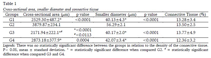
</b>    </font></p> <font face="Verdana" size="2">     ]]></body>
<body><![CDATA[<p>The   histomorphometric analyses of the soleus muscle in relation to the   cross-sectional area, the smaller diameter, and the density of the connective   tissue of the endomysium and the perimysium are presented in <a href="#t1">Table 1</a>. Exercise   (G2) promoted the increase of the cross-sectional area of the fibre in relation   to G1, G3 and G4, [F<sub>(1,20)</sub>=52.14; p&lt;0.0001], as well as increase   the smaller diameter of the fibre in relation to the other groups [F<sub>(1,20)</sub>=49.80;   p&lt;0.0001]. Furthermore, muscle fibre area was higher in G4 compared to G3 [F<sub>(1,20)</sub>=5.190   ;p=0.0338]. There were no changes in connective tissue density, with F<sub>(1,20)</sub>=0.1615; p=0.6920.</p>     <p>&nbsp;</p>    <p align="center"><a name="t1"></a><img src="/img/revistas/mot/v14n1/14n1a05t1.jpg"/></p>    
<p>&nbsp;</p>     <p>
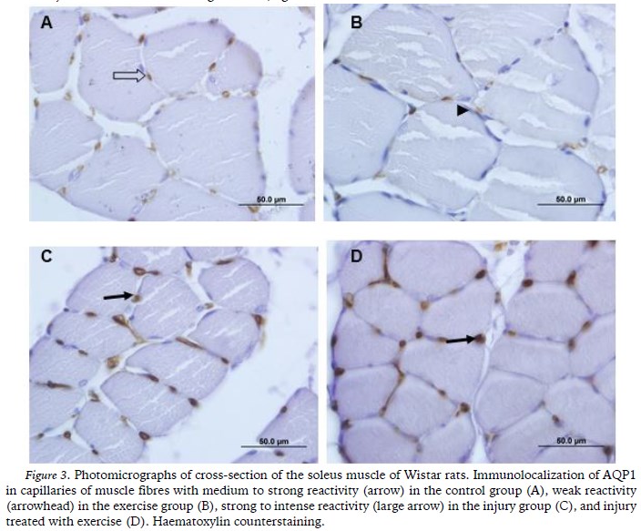
The totals   for blood capillaries, muscle fibres and capillary/fibre ratio for the soleus   muscle are shown in <a href="#t2">Table 2</a>. The injury (G3) was responsible for the increase   of total capillaries in relation G2 and G4 [F<sub>(1,20)</sub>=8.263; p=0.0094].   There was an increase in total muscle fibers per field of analysis in G3 and G4   compared to G2, with F<sub>(1,20)</sub>=14.55; p=0.0011. The capillary/fibber ratio showed no changes between the groups [F<sub>(1,20)</sub>=0.001881; p=0.9658].</p>     <p>&nbsp;</p>    <p align="center"><a name="t2"></a><img src="/img/revistas/mot/v14n1/14n1a05t2.jpg"/></p>    
<p>&nbsp;</p>     <p>
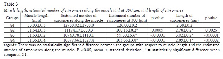
The   analysis of the soleus muscle related to muscle length, estimated number of   sarcomeres along the muscle (and in 300 µm), as well   as the length of the sarcomeres are shown in <a href="#t3">Table 3</a>. There were no   changes in soleus muscle length [F<sub>(1,20)</sub>=0.9356; p=0.3450] and in   the estimated number of sarcomeres along the muscle [F<sub>(1,20)</sub>=1.630;p=0.2164]   between the groups. There was an increase in sarcomere length in G2, G3, G4 when   compared to G1[F<sub>(1,20)</sub>=14.94;p=0.0010]. At the same time, the   estimated of sarcomeres at 300 <i>&#956;</i>m was less in these groups when compared to G1 [F<sub>(1,20)</sub>=16,14;   p=0,0007]. </p>       <p>&nbsp;</p>    ]]></body>
<body><![CDATA[<p align="center"><a name="t3"></a><img src="/img/revistas/mot/v14n1/14n1a05t3.jpg"/></p>    
<p>&nbsp;</p>     <p><b>Morphological and immuno-histochemical analysis of the soleus muscle</b></p>     <p>
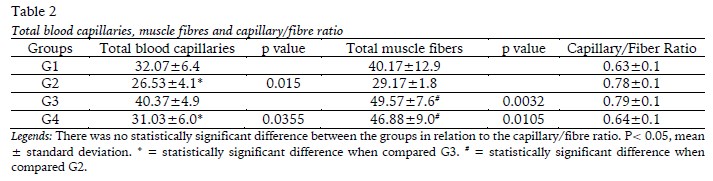
In G1, the   soleus muscle showed normal morphology, with polygonal muscle fibres with   rounded angles and regular contours, myonuclei on the periphery, in the   subsarcolemmal position with fascicular arrangement and with loose connective   tissue in the endomysium and perimysium (<a href="#f1">Figure 1A</a>). In G2, most of the fibres   were hypertrophied compared to the other groups but they maintained a polygonal   shape and fascicular pattern with the nuclei located at the periphery, thereby preserving the muscle structure and organization (<a href="#f1">Figure 1B</a>).</p>     <p>&nbsp;</p>    <p align="center"><a name="f1"></a><img src="/img/revistas/mot/v14n1/14n1a05f1.jpg"/></p>    
<p>&nbsp;</p>     <p>In G3, the   soleus muscle showed signs of damage resulting from denervation. The fibres   were disorganized with irregular contours and centralized nuclei, many of which   showed a circumferential halo and myoblasts in the area of the injury (<a href="#f1">Figure   1C</a>). Signs of degeneration were also observed in the cytoplasm due to the lack   of organization of the myofibrils (<a href="#f1">Figure 1D</a>) and lined-up myoblasts were   visualized at the cell periphery (<a href="#f1">Figure 1E</a>). An increase was observed in the   amount of blood capillaries in the animals in G3, with intense   neovascularization (<a href="#f1">Figure 1E</a>), which was confirmed by the morphometric findings.</p>     <p>In G4,   improvement was observed in the morphology, with the regeneration of the   majority of the muscle fibres, whose characteristics were similar to G1 (<a href="#f1">Figure   1F</a>). However, some fibres with central nuclei and irregular contours were also observed.</p>     <p>
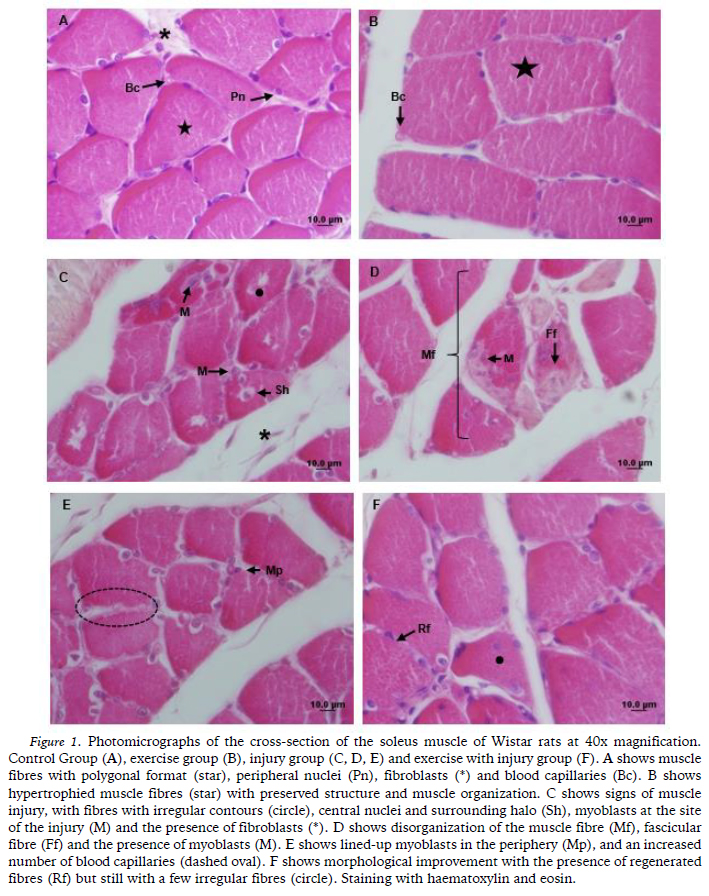
In relation to the density and the structural   organization of the connective tissue in the endomysium and the perimysium,   there were no morphological differences between the groups, all of which were   arranged in typical arrangements with alignment of the fibres and without alterations <a href="#f2">(Figure 2</a>).</p>     ]]></body>
<body><![CDATA[<p>&nbsp;</p>    <p align="center"><a name="f2"></a><img src="/img/revistas/mot/v14n1/14n1a05f2.jpg"/></p>    
<p>&nbsp;</p>     <p>
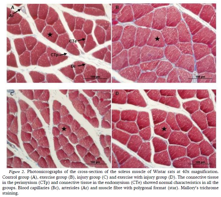
AQP1 was immunolocalized in the endothelium of the   blood capillaries that were present in the muscle fibres of all the groups. The   reactivity was medium to strong in G1 (<a href="#f3">Figure 3A</a>) and weak in G2 (<a href="#f3">Figure 3B</a>).   However, in G3 (<a href="#f3">Figure 3C</a>) and G4 (<a href="#f3">Figure 3D</a>) strong to intense reactivity were observed. </p>     <p>&nbsp;</p>    <p align="center"><a name="f3"></a><img src="/img/revistas/mot/v14n1/14n1a05f3.jpg"/></p>    
<p>&nbsp;</p> </font>     <p>&nbsp;</p>     <p><font size="3" face="Verdana"><b>DISCUSSION</b></font></p> <font face="Verdana" size="2">     <p>The sciatic nerve compression model used in   this study reproduced axonotmesis-type injuries (Silva-Couto et al., 2012),   which results in disruption of neuromuscular communication. The absence of   stimulus changed the morphology of the soleus muscle of the animals in G3,   revealing features that were typical of muscle damage caused by denervation.   Muscle fibres with irregular contours and centralized nuclei were also observed   by Polônio Mazzer, Barbieri, and Matielo-Sverzut (2010) in the tibialis anterior   muscle of rats subjected to a complete section of the sciatic nerve. Salvini Durigan,   Peviani, and Russo (2012)   reported that such structural changes in muscle fibres compromised organ function.</p>     ]]></body>
<body><![CDATA[<p>Blood supply plays an essential role in the   morphological and functional recovery of muscles after nerve injury (Hudlicka,   2011; Wagatsuma et al, 2005). The neovascularization that was observed in G3   enables the supply of nutrients and oxygen that are necessary for tissue   repair. Another study (Camillo, Rocha, &amp; Chopard, 2004), also reported an   increased amount of blood capillaries in the soleus muscle after sectioning of   the sciatic nerve and this characteristic seems to be a response to the   proliferation of endothelial cells that are stimulated by vascular endothelial growth factor (VEGF) (Zhao, Huang, Wu, &amp; Huang 2016).</p>     <p>The sciatic nerve injury in the present study   also promoted changes in the expression of AQP1 in the soleus muscle. AQP1   has been located in red blood cells, the endothelial cells of capillaries,   kidneys, bladder, liver, hippocampus, choroid plexus and other structures.   Although its expression in skeletal muscle is still controversial, there is   agreement about the positive expression of this AQP in the endothelial cells of   muscle capillaries (Wakayama, 2007). Thus, the results regarding the   immunolocalization of AQP1 obtained in the present study were consistent with   the literature because AQP1 was immunolocalized in the endothelium of the soleus muscle of the animals in all of the groups.</p>     <p>The strong to intense reactivity of AQP1 in the   endothelium of the animals from G3 and G4 was a striking result in the present   study and it was evident in an unprecedented manner in the soleus muscle.   Wakayama (2007) argued that the regeneration of skeletal muscle could be   increased by the over-expression of AQP1. That hypothesis is consistent with   our results and it also may be based on other functions of AQPs, such as   facilitating cell migration. Thus, it is believed that the strong reactivity of   AQP1 that was observed in the present study was due to the role of AQP in assisting in the remodelling of the soleus muscle. </p>     <p>Despite the numerous deleterious morphological   characteristics that were observed in the soleus muscle of animals in G3, 21   days after the nerve injury it was possible to observe some signs of muscle   regeneration, such as the presence of centralized nuclei in the fibres, which   was indicative of protein synthesis (Karalaki, Fili,   Philippou, &amp; Koutsilieris,   2009). Tanaka,   Tsubaki, and Tachino (2005) also observed that the soleus muscle recovered spontaneously six weeks after tibial nerve injury.</p>     <p>The plasticity of muscle tissue in response to injury   depends, among other factors, on the functional role of satellite cells   (Karalaki et al., 2009; Wakayama, 2007). These cells are in a quiescent state   in healthy muscles, however, in the case of an injury they proliferate in the   basal lamina, and with the aid of growth factors that act in chemotaxis,   proliferation, and differentiation, the satellite cells form myoblasts. When   they cross the basal lamina the myoblasts release enzymes (trypsin and   pronase), which are capable of dissolving the membrane and reaching the site of   the injury (Carlson, 2014). Evidence suggests that these cells are able to fuse   with muscle fibres in order to repair the injured segment, but they may also   merge to form myotubes, differentiating themselves and originating a new muscle fibre (Wakayama, 2007).</p>     <p>Even though atrophy is frequently observed   after muscle denervation (Russo et al., 2010; Karalaki et al., 2009) this was   not observed in the present study, i.e., there was no decrease in the   cross-sectional area and the diameter of the muscle fibres 21 days after the   injury was produced. The myogenic differentiation factor (myoD) is directly   involved in this process and it increases after denervation because the myoD is   capable of inducing proliferation and differentiation of the satellite cells, thus preventing the appearance of muscular atrophy (Russo et al., 2010).</p>     <p>Different forms of muscle stress can cause   changes in the number of sarcomeres (Ceylan et al., 2014). The exercise   performed in G2 and the nerve damage that was observed in G3 was responsible   for increasing the length of the sarcomeres and concomitantly decreasing their   number at 300 µm. This adjustment occurs so   that there is perfect overlap of the actin and myosin filaments, which allows   the optimal development of tension during contractions (Lima et al., 2007)   thereby showing the adaptability of sarcomeres in the soleus muscle in relation to compressive sciatic nerve injury.</p>     <p>In terms of the animals submitted to resistance   exercise (climbing steps), there were no degenerative morphological changes,   although an increase in the cross-sectional area and the smaller diameter of   the muscle fibres was observed. This hypertrophy was a result of increased   muscle protein synthesis. Without pre-existing injuries, the action of   resistance exercise (climbing steps) has been shown to be effective in inducing muscle hypertrophy in the forelimbs and the hindlimbs (Marqueste et al., 2004).</p>     <p>The potential effect of resistance exercise on   muscle physiology was a determining factor in the morphological improvement of   the soleus muscle of the animals that were subjected to injuries and subsequently   exercised. Aspects of regeneration in the majority of the muscle fibres were   observed in these animals in comparison with animals in G3. Thus, the   resistance exercise (climbing steps), which was started in the acute   post-lesion phase, had a protective effect on the morphology of the soleus   muscle. Salvini et al. (2012) also reported that muscular contraction induced by electrical stimulation has a protective effect on denervated muscles.</p>     <p>The beneficial effects of exercise in   denervation models started at an early stage of reinnervation were also   reported by Marqueste Alliez,   Alluin, Jammes, and Decherchi (2004) who observed that treatment using a treadmill was able to   increase resistance to fatigue, as well as restoring the contractile properties   and mechanosensitivity of the tibialis anterior muscle. A study by Tanaka et   al. (2005) found that the use of a treadmill prevented atrophy of the soleus   muscle after injury to the tibial nerve. These results were attributed to an   increase in protein synthesis as a result of the mechanical stimulation induced   by exercise, and an increase in motor units due to the regeneration of nerve fibres (Betz, Caldwell, &amp; Ribchester, 1980).</p>     ]]></body>
<body><![CDATA[<p>Considering that, to our knowledge, this was   one of the first researches that used as a therapeutic measure the resistance   exercise of stair climbing in the nerve injury, in which the load used was   based on Ilha et al. (2008). We would suggest that further studies should be   performed considering the weight of the animal, the best load, as well as the   intensification of this overload throughout the treatment, so as to have the   maximum possible expression of AQP1 and that this may determine the acceleration of recovery.</p> </font>     <p>&nbsp;</p>     <p><font size="3" face="Verdana"><b>CONCLUSION</b></font><font face="Verdana" size="2">    </font></p> <font face="Verdana" size="2">     <p>Compression of the sciatic nerve was capable of   promoting the effects of muscle injury and AQP1 was   immunolocalized in the endothelium of blood capillaries, and 21 days after   axonotmesis of the soleus muscle the intrinsic potential for recovery was   evident. It is   important to start resistance exercise in the acute phase after denervation   because the resistance exercise (climbing steps) acted as a protective factor in regeneration.</p>     <p>&nbsp;</p> </font>     <p><font size="3" face="Verdana"><b>REFERENCES</b></font></p> <font face="Verdana" size="2">     <!-- ref --><p>Artifon,   E. L., Silva, L. I., Ribeiro, L. F. C., Brancalhão, R. M. C., &amp; Bertolini,   G. R. F. (2013). Treinamento aeróbico prévio à compressão nervosa: análise da   morfometria muscular de ratos. <i>Revista Brasileira     de Medicina do Esporte,</i> <i>19</i>(1), 66-69. doi: 0.1590/S1517-86922013000100014&nbsp;    &nbsp;&nbsp;&nbsp;&nbsp;&nbsp;&nbsp;&nbsp;&nbsp;[&#160;<a href="javascript:void(0);" onclick="javascript: window.open('/scielo.php?script=sci_nlinks&ref=373934&pid=S1646-107X201800010000500001&lng=','','width=640,height=500,resizable=yes,scrollbars=1,menubar=yes,');">Links</a>&#160;]<!-- end-ref --></p>     <!-- ref --><p>Betz, W. J., Caldwell, J. H., &amp; Ribchester,   R. R. (1980). The effects of partial denervation at birth on the development of muscle   fibres and motor units in rat lumbrical muscle. <i>Journal of Physiology,</i> <i>303</i>, 265-279.    &nbsp;&nbsp;&nbsp;&nbsp;&nbsp;&nbsp;&nbsp;&nbsp;[&#160;<a href="javascript:void(0);" onclick="javascript: window.open('/scielo.php?script=sci_nlinks&ref=373936&pid=S1646-107X201800010000500002&lng=','','width=640,height=500,resizable=yes,scrollbars=1,menubar=yes,');">Links</a>&#160;]<!-- end-ref --></p>     ]]></body>
<body><![CDATA[<!-- ref --><p>Bosi,   P. L., Delfino, G. B., Durigan, J. L. Q., Cancelliero, K. M., Polacow, M. L. O,   &amp; Silva, C. A. (2008). Metformina   minimiza as alterações morfométricas no músculo sóleo de ratos submetidos à   imobilização articular. <i>Revista     Brasileira de Medicina do Esporte</i>, <i>14</i>(5), 436-439. doi: 10.1590/S1517-86922008000500007&nbsp;    &nbsp;&nbsp;&nbsp;&nbsp;&nbsp;&nbsp;&nbsp;&nbsp;[&#160;<a href="javascript:void(0);" onclick="javascript: window.open('/scielo.php?script=sci_nlinks&ref=373938&pid=S1646-107X201800010000500003&lng=','','width=640,height=500,resizable=yes,scrollbars=1,menubar=yes,');">Links</a>&#160;]<!-- end-ref --></p>     <!-- ref --><p>Caierão,   Q. M., Betini, J., Teodori, R. M., &amp; Minamoto, V. B. (2008). O efeito do intervalo da estimulação elétrica no músculo desnervado de   rato. <i>Revista Brasileira de Fisioterapia</i>, <i>12</i>(2), 143-148. doi:10.1590/S1413-35552008000200011&nbsp;    &nbsp;&nbsp;&nbsp;&nbsp;&nbsp;&nbsp;&nbsp;&nbsp;[&#160;<a href="javascript:void(0);" onclick="javascript: window.open('/scielo.php?script=sci_nlinks&ref=373940&pid=S1646-107X201800010000500004&lng=','','width=640,height=500,resizable=yes,scrollbars=1,menubar=yes,');">Links</a>&#160;]<!-- end-ref --></p>     <!-- ref --><p>Câmera,   C. N. S., Brito, M. V. H., Silveira, E. L., Silva, D. S. G., Simões, V. R. F,   &amp; Pontes, R. W. F. (2011). Histological analysis of low-intensity laser therapy effects in peripheral nerve regeneration in Wistar rats. <i>Acta Cirurgica Brasileira</i>, <i>26</i>(1), 12-18. doi: 10.1590/S0102-86502011000100004&nbsp;    &nbsp;&nbsp;&nbsp;&nbsp;&nbsp;&nbsp;&nbsp;&nbsp;[&#160;<a href="javascript:void(0);" onclick="javascript: window.open('/scielo.php?script=sci_nlinks&ref=373942&pid=S1646-107X201800010000500005&lng=','','width=640,height=500,resizable=yes,scrollbars=1,menubar=yes,');">Links</a>&#160;]<!-- end-ref --></p>     <!-- ref --><p>Camillo,   A. C., Rocha, R. C., &amp; Chopard, R. P. (2004). Structural and microvascular study of soleous muscle of wistar rats after section of the sciatic nerve. <i>Arquivos de Neuro-Psiquiatria,</i> <i>62</i>(3), 835-838. doi: 10.1590/S0004-282X2004000500018&nbsp;    &nbsp;&nbsp;&nbsp;&nbsp;&nbsp;&nbsp;&nbsp;&nbsp;[&#160;<a href="javascript:void(0);" onclick="javascript: window.open('/scielo.php?script=sci_nlinks&ref=373944&pid=S1646-107X201800010000500006&lng=','','width=640,height=500,resizable=yes,scrollbars=1,menubar=yes,');">Links</a>&#160;]<!-- end-ref --></p>     <!-- ref --><p>Cassilhas,   R. C., Reis, I. T., Venâncio, D., Fernandes, J., Tufik, S., &amp; Mello, M. T.   (2013). Animal   model for progressive resistance exercise: a detailed description of model and   its implications for basic research in exercise. <i>Revista Motriz,</i> <i>19</i>(1), 178-184. doi: 10.1590/S1980-65742013000100018&nbsp;    &nbsp;&nbsp;&nbsp;&nbsp;&nbsp;&nbsp;&nbsp;&nbsp;[&#160;<a href="javascript:void(0);" onclick="javascript: window.open('/scielo.php?script=sci_nlinks&ref=373946&pid=S1646-107X201800010000500007&lng=','','width=640,height=500,resizable=yes,scrollbars=1,menubar=yes,');">Links</a>&#160;]<!-- end-ref --></p>     ]]></body>
<body><![CDATA[<!-- ref --><p>Carlson, B. M. (2014). The biology of long-term denervated skeletal muscle. <i>European Journal Translational Myology – BAM, 24</i>(1), 5-11. doi: 10.4081/ejtm.2014.3293&nbsp;&nbsp;&nbsp;&nbsp;&nbsp;&nbsp;&nbsp;&nbsp;[&#160;<a href="javascript:void(0);" onclick="javascript: window.open('/scielo.php?script=sci_nlinks&ref=373948&pid=S1646-107X201800010000500008&lng=','','width=640,height=500,resizable=yes,scrollbars=1,menubar=yes,');">Links</a>&#160;]<!-- end-ref --><!-- ref --><p>Ceylan,   O., Seyfettinoglu, F., Dulgeroglu, A. M., Avci, A., Bayram, B., &amp; Bora, O.   (2014). A. Histomorphological comparison of immobilization and desnervation   atrophies. <i>Acta Orthopaedica et     Traumatologica Túrcica,</i> <i>48</i>(3), 320-325. doi: 10.3944/AOTT.2014.2993&nbsp;&nbsp;&nbsp;&nbsp;&nbsp;&nbsp;&nbsp;&nbsp;[&#160;<a href="javascript:void(0);" onclick="javascript: window.open('/scielo.php?script=sci_nlinks&ref=373949&pid=S1646-107X201800010000500009&lng=','','width=640,height=500,resizable=yes,scrollbars=1,menubar=yes,');">Links</a>&#160;]<!-- end-ref --><!-- ref --><p>Cheng, F. C., Sheu, M. L., Su,   H. L., Chen, Y. J., Chen, C. J., Chiu, W.T, Sheehan. J., &amp; Pan, H. C.   (2013). The effect of exercise on mobilization of hematopoietic progenitor   cells involved in the repair of sciatic nerve crush injury. <i>Journal of Neurosurgery,</i> <i>118</i>(3), 594-605. doi: 10.3171/2012.8.JNS111580&nbsp;&nbsp;&nbsp;&nbsp;&nbsp;&nbsp;&nbsp;&nbsp;[&#160;<a href="javascript:void(0);" onclick="javascript: window.open('/scielo.php?script=sci_nlinks&ref=373950&pid=S1646-107X201800010000500010&lng=','','width=640,height=500,resizable=yes,scrollbars=1,menubar=yes,');">Links</a>&#160;]<!-- end-ref --><!-- ref --><p>Coutinho,   E. L., Gomes, A. R. S., França, C. N., Oishi, J., &amp; Salvini, T. F. (2004).   Effect of passive stretching on the immobilized soleus muscle fiber morphology<i>. Brazilian Journal of Medicine and Biology     and Research</i>, <i>37</i>(12), 1853-1861. doi: 10.1590/S0100-879X2004001200011&nbsp;    &nbsp;&nbsp;&nbsp;&nbsp;&nbsp;&nbsp;&nbsp;&nbsp;[&#160;<a href="javascript:void(0);" onclick="javascript: window.open('/scielo.php?script=sci_nlinks&ref=373951&pid=S1646-107X201800010000500011&lng=','','width=640,height=500,resizable=yes,scrollbars=1,menubar=yes,');">Links</a>&#160;]<!-- end-ref --></p>     <!-- ref --><p>Fernandes,   T., Roque, F. R., Magalhães, F. C., Carmo, E. C., &amp; Oliveira, E. M. (2012).   O treinamento físico aeróbio corrige a rarefação capilar e as alterações nas   proporções dos tipos de fibra muscular esquelética em ratos espontaneamente   hipertensos. <i>Revista Brasileira de Medicina do Esporte</i>, <i>18</i>(4), 267-272. doi: 10.1590/S1517-86922012000400010&nbsp;    &nbsp;&nbsp;&nbsp;&nbsp;&nbsp;&nbsp;&nbsp;&nbsp;[&#160;<a href="javascript:void(0);" onclick="javascript: window.open('/scielo.php?script=sci_nlinks&ref=373953&pid=S1646-107X201800010000500012&lng=','','width=640,height=500,resizable=yes,scrollbars=1,menubar=yes,');">Links</a>&#160;]<!-- end-ref --></p>     <!-- ref --><p>Frasson,   N. F. V., Taciro, C., &amp; Parizotto, N. A. (2009). Análise nanoestrutural da   ação do ultra-som terapêutico sobre o processo de regeneração do tendão de   ratos. <i>Revista Fisioterapia e Pesquisa, 16</i>(3),   198-204. doi: 10.1590/S1809-29502009000300002&nbsp;    &nbsp;&nbsp;&nbsp;&nbsp;&nbsp;&nbsp;&nbsp;&nbsp;[&#160;<a href="javascript:void(0);" onclick="javascript: window.open('/scielo.php?script=sci_nlinks&ref=373955&pid=S1646-107X201800010000500013&lng=','','width=640,height=500,resizable=yes,scrollbars=1,menubar=yes,');">Links</a>&#160;]<!-- end-ref --></p>     <!-- ref --><p>Gaffuri, J., Meireles, A., Rocha, B. P., Rosa, C. T.,   Artifon, E.L., Silva, L. I., Moreira, N.    B., &amp; Bertolini, G. R. F. (2011). Avaliação do exercício físico como   fator de analgesia em um modelo experimental de ciatalgia. <i>Revista Brasileira de Medicina do Esporte,</i> <i>17</i>(2), 115-118. doi: 10.1590/S1517-86922011000200009&nbsp;    &nbsp;&nbsp;&nbsp;&nbsp;&nbsp;&nbsp;&nbsp;&nbsp;[&#160;<a href="javascript:void(0);" onclick="javascript: window.open('/scielo.php?script=sci_nlinks&ref=373957&pid=S1646-107X201800010000500014&lng=','','width=640,height=500,resizable=yes,scrollbars=1,menubar=yes,');">Links</a>&#160;]<!-- end-ref --></p>     <!-- ref --><p>Gorio, A., Carmignotto, G., Finesso, M., Polato,   P., &amp; Nunzi, M. G. (1983). Muscle reinnervation – II. Sprouting,   synapse formation and repression. <i>Neuroscience,</i> <i>8</i>(3), 403-416. doi: 10.1016/0306-4522(83)90188-4&nbsp;&nbsp;&nbsp;&nbsp;&nbsp;&nbsp;&nbsp;&nbsp;[&#160;<a href="javascript:void(0);" onclick="javascript: window.open('/scielo.php?script=sci_nlinks&ref=373959&pid=S1646-107X201800010000500015&lng=','','width=640,height=500,resizable=yes,scrollbars=1,menubar=yes,');">Links</a>&#160;]<!-- end-ref --><!-- ref --><p>Hornberger,   T. A. J., &amp; Farrar, R. P. (2004). Physiological hypertrophy of the FHL muscle following 8 weeks of progressive resistance exercise in the rat. <i>Canadian Journal of applied physiology</i>, <i>29</i>(1), 16-31. doi: 10.1139/h04-002&nbsp;&nbsp;&nbsp;&nbsp;&nbsp;&nbsp;&nbsp;&nbsp;[&#160;<a href="javascript:void(0);" onclick="javascript: window.open('/scielo.php?script=sci_nlinks&ref=373960&pid=S1646-107X201800010000500016&lng=','','width=640,height=500,resizable=yes,scrollbars=1,menubar=yes,');">Links</a>&#160;]<!-- end-ref --><!-- ref --><p>Hudlicka, O. (2011). Microcirculation in skeletal muscle. <i>Muscle, Ligaments and Tendons Journal,</i> <i>1</i>(1), 3-11.    &nbsp;&nbsp;&nbsp;&nbsp;&nbsp;&nbsp;&nbsp;&nbsp;[&#160;<a href="javascript:void(0);" onclick="javascript: window.open('/scielo.php?script=sci_nlinks&ref=373961&pid=S1646-107X201800010000500017&lng=','','width=640,height=500,resizable=yes,scrollbars=1,menubar=yes,');">Links</a>&#160;]<!-- end-ref --></p>     <!-- ref --><p>Ilha,   J., Araujo, R. T., Malysz, T., Hermel, E. E., Rigon, P., Xavier, L. L., &amp;   Achaval, M. (2008). Endurance and resistance exercise training programs elicit specific   effects on sciatic nerve regeneration after experimental traumatic lesion in   rats<i>. Neurorehabilitation     and Neural Repair</i>, <i>22</i>(4), 355-366. doi: 10.1177/1545968307313502&nbsp;&nbsp;&nbsp;&nbsp;&nbsp;&nbsp;&nbsp;&nbsp;[&#160;<a href="javascript:void(0);" onclick="javascript: window.open('/scielo.php?script=sci_nlinks&ref=373963&pid=S1646-107X201800010000500018&lng=','','width=640,height=500,resizable=yes,scrollbars=1,menubar=yes,');">Links</a>&#160;]<!-- end-ref --><!-- ref --><p>Kiernan, J. (2015). <i>Histological   and Histochemical Methods: Theory and Practice</i> (5<sup>th</sup> Ed.). Banbury: Scion Publishing Limited.    &nbsp;&nbsp;&nbsp;&nbsp;&nbsp;&nbsp;&nbsp;&nbsp;[&#160;<a href="javascript:void(0);" onclick="javascript: window.open('/scielo.php?script=sci_nlinks&ref=373964&pid=S1646-107X201800010000500019&lng=','','width=640,height=500,resizable=yes,scrollbars=1,menubar=yes,');">Links</a>&#160;]<!-- end-ref --></p>     <!-- ref --><p> Junior, G. M. R.,   Bueno, C. R. S., Heubel, A., Bortoluci, C. H. F., Simionato, L. H., Daré, L.   R., Silva, M. P., Dias, D. V. (2013). Efeito da corrente alternada simétrica sinusoidal na musculatura estriada esquelética desnervada experimentalmente. <i>Salusvita,</i> <i>32</i>(3), 211-225.    &nbsp;&nbsp;&nbsp;&nbsp;&nbsp;&nbsp;&nbsp;&nbsp;[&#160;<a href="javascript:void(0);" onclick="javascript: window.open('/scielo.php?script=sci_nlinks&ref=373966&pid=S1646-107X201800010000500020&lng=','','width=640,height=500,resizable=yes,scrollbars=1,menubar=yes,');">Links</a>&#160;]<!-- end-ref --></p>     ]]></body>
<body><![CDATA[<!-- ref --><p>Karalaki,   M., Fili, S., Philippou, A., &amp; Koutsilieris, M. (2009). Muscle   Regeneration: Cellular and Molecular Events<i>. In vivo,</i> <i>23</i>(5), 779-796.    &nbsp;&nbsp;&nbsp;&nbsp;&nbsp;&nbsp;&nbsp;&nbsp;[&#160;<a href="javascript:void(0);" onclick="javascript: window.open('/scielo.php?script=sci_nlinks&ref=373968&pid=S1646-107X201800010000500021&lng=','','width=640,height=500,resizable=yes,scrollbars=1,menubar=yes,');">Links</a>&#160;]<!-- end-ref --></p>     <!-- ref --><p>Kitchen,   P., Day, R. E., Salman, M. M., Conner, M.T., Bill, R. M., &amp; Conner, A. C.   (2015). Beyond water homeostasis: diverse functional roles of mammalian   aquaporins. <i>Biochimica et Biophysica Acta,</i> <i>1850</i>(12), 2410-2421. doi:10.1016/j.bbagen.2015.08.023&nbsp;&nbsp;&nbsp;&nbsp;&nbsp;&nbsp;&nbsp;&nbsp;[&#160;<a href="javascript:void(0);" onclick="javascript: window.open('/scielo.php?script=sci_nlinks&ref=373970&pid=S1646-107X201800010000500022&lng=','','width=640,height=500,resizable=yes,scrollbars=1,menubar=yes,');">Links</a>&#160;]<!-- end-ref --><!-- ref --><p>Kobayashi,   S., Yoshizawa, H., &amp; Yamada, S. (2004). Pathology   of lumbar nerve root compression. Part 2: morphological and immunohistochemical   changes of dorsal root ganglion. <i>Journal Orthopaedic Research,</i> <i>22</i>(1), 180-188. doi: 10.1016/S0736-0266(03)00132-3&nbsp;&nbsp;&nbsp;&nbsp;&nbsp;&nbsp;&nbsp;&nbsp;[&#160;<a href="javascript:void(0);" onclick="javascript: window.open('/scielo.php?script=sci_nlinks&ref=373971&pid=S1646-107X201800010000500023&lng=','','width=640,height=500,resizable=yes,scrollbars=1,menubar=yes,');">Links</a>&#160;]<!-- end-ref --><!-- ref --><p>Li, R., Liu, Z., Pan, Y., Chen, L., Zhang, Z., &amp; Lu, L. (2014). Peripheral nerve injuries treatment: a systematic review. <i>Cell Biochemistry and Biophysics.</i> <i>68</i>(3). 449-454. doi: 10.1007/s12013-013-9742-1&nbsp;&nbsp;&nbsp;&nbsp;&nbsp;&nbsp;&nbsp;&nbsp;[&#160;<a href="javascript:void(0);" onclick="javascript: window.open('/scielo.php?script=sci_nlinks&ref=373972&pid=S1646-107X201800010000500024&lng=','','width=640,height=500,resizable=yes,scrollbars=1,menubar=yes,');">Links</a>&#160;]<!-- end-ref --><!-- ref --><p>Lima,   S. C., Caierão, Q. M., Durigan, J. L. Q., Schwarzenbeck, A., Silva, C. A,   Minamoto, V. B, &amp; Guirro, R. R. J. (2007). Curto período de imobilização provoca alterações morfométricas e mecânicas no músculo de rato. <i>Revista Brasileira de Fisioterapia,</i> <i>11</i>(4), 297-302.    &nbsp;&nbsp;&nbsp;&nbsp;&nbsp;&nbsp;&nbsp;&nbsp;[&#160;<a href="javascript:void(0);" onclick="javascript: window.open('/scielo.php?script=sci_nlinks&ref=373973&pid=S1646-107X201800010000500025&lng=','','width=640,height=500,resizable=yes,scrollbars=1,menubar=yes,');">Links</a>&#160;]<!-- end-ref --></p>     <!-- ref --><p>Lim,   J. H., Kim, D. H., Han, D. W., Kwak, J. Y., Bae, H. R. (2016). The effect of AQP3   deficiency on fuel selection during bout of exhausting exercise. <i>Pflugers Arch. – European. Journal of Physiology</i>, <i>468</i>, 1283-1293. doi: 10.1007/s00424-016-1827-4.    &nbsp;&nbsp;&nbsp;&nbsp;&nbsp;&nbsp;&nbsp;&nbsp;[&#160;<a href="javascript:void(0);" onclick="javascript: window.open('/scielo.php?script=sci_nlinks&ref=373975&pid=S1646-107X201800010000500026&lng=','','width=640,height=500,resizable=yes,scrollbars=1,menubar=yes,');">Links</a>&#160;]<!-- end-ref --></p>     <!-- ref --><p>Marqueste, T., Alliez, J. R., Alluin, O., Jammes,   Y., &amp; Decherchi, P. (2004). Neuromuscular rehabilitation by treadmill running or electrical stimulation after peripheral nerve injury and repair. <i>Journal of Applied Physiology,</i> <i>96</i>(5), 1988-1995. doi: 10.1152/japplphysiol.00775.2003&nbsp;    &nbsp;&nbsp;&nbsp;&nbsp;&nbsp;&nbsp;&nbsp;&nbsp;[&#160;<a href="javascript:void(0);" onclick="javascript: window.open('/scielo.php?script=sci_nlinks&ref=373977&pid=S1646-107X201800010000500027&lng=','','width=640,height=500,resizable=yes,scrollbars=1,menubar=yes,');">Links</a>&#160;]<!-- end-ref --></p>     <!-- ref --><p>Moore, K. L., &amp; Dalley, A. F. (2007). <i>Anatomia Orientada para a Clínica</i>. Rio de Janeiro: Guanabara Koogan.    &nbsp;&nbsp;&nbsp;&nbsp;&nbsp;&nbsp;&nbsp;&nbsp;[&#160;<a href="javascript:void(0);" onclick="javascript: window.open('/scielo.php?script=sci_nlinks&ref=373979&pid=S1646-107X201800010000500028&lng=','','width=640,height=500,resizable=yes,scrollbars=1,menubar=yes,');">Links</a>&#160;]<!-- end-ref --></p>     <!-- ref --><p>Polônio,   J. T., Mazzer, N., Barbieri, C. H., &amp; Matielo-Sverzut, A. C. (2010).   Eletroestimulação seletiva mantem estrutura e função do tibial desnervado de ratos. <i>Acta Ortopédica Brasileira</i>, <i>18</i>(2), 85-89. doi: 10.1590/S1413-78522010000200005&nbsp;    &nbsp;&nbsp;&nbsp;&nbsp;&nbsp;&nbsp;&nbsp;&nbsp;[&#160;<a href="javascript:void(0);" onclick="javascript: window.open('/scielo.php?script=sci_nlinks&ref=373981&pid=S1646-107X201800010000500029&lng=','','width=640,height=500,resizable=yes,scrollbars=1,menubar=yes,');">Links</a>&#160;]<!-- end-ref --></p>     <!-- ref --><p>Rivera, M. A., Martinez, J. L, Carrion, A., &amp; Fahey   T. D. (2011). AQP-1 association with body fluid loss in   10-km runners. <i>International Journal Sports of Medicine</i>, <i>32</i>, 229-233. doi: 10.1055/s-0030-1268489 &nbsp;&nbsp;&nbsp;&nbsp;&nbsp;&nbsp;&nbsp;&nbsp;[&#160;<a href="javascript:void(0);" onclick="javascript: window.open('/scielo.php?script=sci_nlinks&ref=373983&pid=S1646-107X201800010000500030&lng=','','width=640,height=500,resizable=yes,scrollbars=1,menubar=yes,');">Links</a>&#160;]<!-- end-ref --><!-- ref --><p>Russo, T. L., Peviani, S. M., Durigan, J. L.Q,   Gigo-Benato, D., Delfino, G. B., &amp; Salvini, T. F. (2010). Stretching and electrical stimulation reduce the accumulation of MyoD, myostatin and atrogin-1 in denervated rat skeletal muscle<i>. Journal of Muscle Research and cell motility,</i> <i>31</i>(1), 45-57. doi: 10.1007/s10974-010-9203-z&nbsp;&nbsp;&nbsp;&nbsp;&nbsp;&nbsp;&nbsp;&nbsp;[&#160;<a href="javascript:void(0);" onclick="javascript: window.open('/scielo.php?script=sci_nlinks&ref=373984&pid=S1646-107X201800010000500031&lng=','','width=640,height=500,resizable=yes,scrollbars=1,menubar=yes,');">Links</a>&#160;]<!-- end-ref --><!-- ref --><p>Salvini,   T. F., Durigan, J. L. Q., Peviani, S. M., &amp; Russo, T. L. (2012). Efeitos da   eletroestimulação e do alongamento muscular sobre a adaptação do músculo desnervado – implicações para a fisioterapia. <i>Revista Brasileira de Fisioterapia,</i> <i>16</i>(3), 175-183. doi: 10.1590/S1413-35552012005000027&nbsp;    &nbsp;&nbsp;&nbsp;&nbsp;&nbsp;&nbsp;&nbsp;&nbsp;[&#160;<a href="javascript:void(0);" onclick="javascript: window.open('/scielo.php?script=sci_nlinks&ref=373985&pid=S1646-107X201800010000500032&lng=','','width=640,height=500,resizable=yes,scrollbars=1,menubar=yes,');">Links</a>&#160;]<!-- end-ref --></p>     <!-- ref --><p>Sebben,   A. D., Cocolichio, F., Schmitt, A. P. V., Curra, M. D., Silva, P. V., Tres, G.   L., &amp; Silva, J. B. (2011). Efeitos de fatores neurotróficos sobre o reparo   de nervo periférico. <i>Scientia Médica</i>, <i>21</i>(2), 81-89.    &nbsp;&nbsp;&nbsp;&nbsp;&nbsp;&nbsp;&nbsp;&nbsp;[&#160;<a href="javascript:void(0);" onclick="javascript: window.open('/scielo.php?script=sci_nlinks&ref=373987&pid=S1646-107X201800010000500033&lng=','','width=640,height=500,resizable=yes,scrollbars=1,menubar=yes,');">Links</a>&#160;]<!-- end-ref --></p>     <!-- ref --><p>Silva-Couto,   M. A., Gigo-Benato, D., Tim, C. R., Parizotto, N. A., Salvini T. F., &amp;   Russo, T. L. (2012). Effects of low-level laser therapy after nerve reconstruction in rat   denervated soleus muscle adaptation. <i>Revista Brasileira de Fisioterapia</i>, <i>16</i>(4), 320-327.    &nbsp;&nbsp;&nbsp;&nbsp;&nbsp;&nbsp;&nbsp;&nbsp;[&#160;<a href="javascript:void(0);" onclick="javascript: window.open('/scielo.php?script=sci_nlinks&ref=373989&pid=S1646-107X201800010000500034&lng=','','width=640,height=500,resizable=yes,scrollbars=1,menubar=yes,');">Links</a>&#160;]<!-- end-ref --></p>     <!-- ref --><p>Silva,   L. I., Meirelhes, A., Nascimento, C. M., Rocha, B. P., Rosa, C. T., Ribeiro, L.   F. C., Brancalhão, R. M. C., &amp; Bertolini, G. R. F. (2013). Avaliação de   parâmetros histomorfométricos em sóleos de ratos submetidos à remobilização por   salto em meio aquático. <i>Revista     Brasileira de Medicina do Esporte</i>, <i>19</i>(3), 219-222.    &nbsp;&nbsp;&nbsp;&nbsp;&nbsp;&nbsp;&nbsp;&nbsp;[&#160;<a href="javascript:void(0);" onclick="javascript: window.open('/scielo.php?script=sci_nlinks&ref=373991&pid=S1646-107X201800010000500035&lng=','','width=640,height=500,resizable=yes,scrollbars=1,menubar=yes,');">Links</a>&#160;]<!-- end-ref --></p>     <!-- ref --><p>Tanaka, S., Tsubaki, A., &amp; Tachino, K. (2005). Effect of exercise training after partial denervation in rat soleus   muscles. <i>Journal of Physical Therapy Science,</i> <i>17</i>(2), 97-101. doi: 10.1589/jpts.17.97&nbsp;&nbsp;&nbsp;&nbsp;&nbsp;&nbsp;&nbsp;&nbsp;[&#160;<a href="javascript:void(0);" onclick="javascript: window.open('/scielo.php?script=sci_nlinks&ref=373993&pid=S1646-107X201800010000500036&lng=','','width=640,height=500,resizable=yes,scrollbars=1,menubar=yes,');">Links</a>&#160;]<!-- end-ref --><!-- ref --><p>Wagatsuma, A., Tamaki, H., &amp; Ogita, F. (2005).   Capillary supply and gene expression of angiogenesis-related factors in murine   skeletal muscle following denervation. <i>Experimental     Physiology</i>, <i>90</i>(3), 403-409. doi: 10.1113/exoohysiol.2004.029769&nbsp;&nbsp;&nbsp;&nbsp;&nbsp;&nbsp;&nbsp;&nbsp;[&#160;<a href="javascript:void(0);" onclick="javascript: window.open('/scielo.php?script=sci_nlinks&ref=373994&pid=S1646-107X201800010000500037&lng=','','width=640,height=500,resizable=yes,scrollbars=1,menubar=yes,');">Links</a>&#160;]<!-- end-ref --><!-- ref --><p>Wakayama,   Y. (2007). Skeletal muscle regeneration may be enhanced by overexpression of   aquaporin 1 in intramuscular capillary endothelial cells. <i>Medical Hypotheses,</i> <i>68</i>, 856-859. doi: 10.1016/j.mehy.2006.07.056&nbsp;&nbsp;&nbsp;&nbsp;&nbsp;&nbsp;&nbsp;&nbsp;[&#160;<a href="javascript:void(0);" onclick="javascript: window.open('/scielo.php?script=sci_nlinks&ref=373995&pid=S1646-107X201800010000500038&lng=','','width=640,height=500,resizable=yes,scrollbars=1,menubar=yes,');">Links</a>&#160;]<!-- end-ref --><!-- ref --><p>Zhao, H., Huang, H., Wu, J., &amp; Huang, P. (2016).   Specialization of mitochondrial and vascular oxidant modulated VEGFR in the   denervated skeletal muscle<i>. Cell     Signalling,</i> <i>25</i>(11), 2106-2114. doi: 10.1016/j.cellsig.2013.06.014.    &nbsp;&nbsp;&nbsp;&nbsp;&nbsp;&nbsp;&nbsp;&nbsp;[&#160;<a href="javascript:void(0);" onclick="javascript: window.open('/scielo.php?script=sci_nlinks&ref=373996&pid=S1646-107X201800010000500039&lng=','','width=640,height=500,resizable=yes,scrollbars=1,menubar=yes,');">Links</a>&#160;]<!-- end-ref --></p>     ]]></body>
<body><![CDATA[<p>&nbsp;</p> </font>     <p><font size="2" face="Verdana"><b>Acknowledgments:</b>    <br>   Nothing to declare<b>    <br>   Conflict of interests:</b>    <br>   Nothing to declare.<b>    <br>   Funding:    <br>   </b></font><font face="Verdana"><b></b><font size="2">Nothing to declare.</font></font></p>     <p><font size="2" face="Verdana">Manuscript received at March  29th 2017; Accepted at September 5th 2017 </font></p>     <p>&nbsp;</p>     <p>&nbsp;</p> <font size="2" face="Verdana"><i><a name="end"></a></i><a href="#top">Correspondence to:</a> Rua Universit&aacute;ria, 2069, 85819-110 Cascavel, PR, Brazil. <i>E-mail</i>: <a href="mailto:keli.lovison@hotmail.com">keli.lovison@hotmail.com</a> </font>     ]]></body>
<body><![CDATA[ ]]></body><back>
<ref-list>
<ref id="B1">
<nlm-citation citation-type="journal">
<person-group person-group-type="author">
<name>
<surname><![CDATA[Artifon]]></surname>
<given-names><![CDATA[E. L.]]></given-names>
</name>
<name>
<surname><![CDATA[Silva]]></surname>
<given-names><![CDATA[L. I.]]></given-names>
</name>
<name>
<surname><![CDATA[Ribeiro]]></surname>
<given-names><![CDATA[L. F. C.]]></given-names>
</name>
<name>
<surname><![CDATA[Brancalhão]]></surname>
<given-names><![CDATA[R. M. C.]]></given-names>
</name>
<name>
<surname><![CDATA[Bertolini]]></surname>
<given-names><![CDATA[G. R. F.]]></given-names>
</name>
</person-group>
<article-title xml:lang="pt"><![CDATA[Treinamento aeróbico prévio à compressão nervosa: análise da morfometria muscular de ratos]]></article-title>
<source><![CDATA[Revista Brasileira de Medicina do Esporte]]></source>
<year>2013</year>
<volume>19</volume>
<numero>1</numero>
<issue>1</issue>
<page-range>66-69</page-range></nlm-citation>
</ref>
<ref id="B2">
<nlm-citation citation-type="journal">
<person-group person-group-type="author">
<name>
<surname><![CDATA[Betz]]></surname>
<given-names><![CDATA[W. J.]]></given-names>
</name>
<name>
<surname><![CDATA[Caldwell]]></surname>
<given-names><![CDATA[J. H.]]></given-names>
</name>
<name>
<surname><![CDATA[Ribchester]]></surname>
<given-names><![CDATA[R. R.]]></given-names>
</name>
</person-group>
<article-title xml:lang="en"><![CDATA[The effects of partial denervation at birth on the development of muscle fibres and motor units in rat lumbrical muscle]]></article-title>
<source><![CDATA[Journal of Physiology]]></source>
<year>1980</year>
<volume>303</volume>
<page-range>265-279</page-range></nlm-citation>
</ref>
<ref id="B3">
<nlm-citation citation-type="journal">
<person-group person-group-type="author">
<name>
<surname><![CDATA[Bosi]]></surname>
<given-names><![CDATA[P. L.]]></given-names>
</name>
<name>
<surname><![CDATA[Delfino]]></surname>
<given-names><![CDATA[G. B.]]></given-names>
</name>
<name>
<surname><![CDATA[Durigan]]></surname>
<given-names><![CDATA[J. L. Q.]]></given-names>
</name>
<name>
<surname><![CDATA[Cancelliero]]></surname>
<given-names><![CDATA[K. M.]]></given-names>
</name>
<name>
<surname><![CDATA[Polacow]]></surname>
<given-names><![CDATA[M. L. O]]></given-names>
</name>
<name>
<surname><![CDATA[Silva]]></surname>
<given-names><![CDATA[C. A.]]></given-names>
</name>
</person-group>
<article-title xml:lang="pt"><![CDATA[Metformina minimiza as alterações morfométricas no músculo sóleo de ratos submetidos à imobilização articular]]></article-title>
<source><![CDATA[Revista Brasileira de Medicina do Esporte]]></source>
<year>2008</year>
<volume>14</volume>
<numero>5</numero>
<issue>5</issue>
<page-range>436-439</page-range></nlm-citation>
</ref>
<ref id="B4">
<nlm-citation citation-type="journal">
<person-group person-group-type="author">
<name>
<surname><![CDATA[Caierão]]></surname>
<given-names><![CDATA[Q. M.]]></given-names>
</name>
<name>
<surname><![CDATA[Betini]]></surname>
<given-names><![CDATA[J.]]></given-names>
</name>
<name>
<surname><![CDATA[Teodori]]></surname>
<given-names><![CDATA[R. M.]]></given-names>
</name>
<name>
<surname><![CDATA[Minamoto]]></surname>
<given-names><![CDATA[V. B.]]></given-names>
</name>
</person-group>
<article-title xml:lang="pt"><![CDATA[O efeito do intervalo da estimulação elétrica no músculo desnervado de rato]]></article-title>
<source><![CDATA[Revista Brasileira de Fisioterapia]]></source>
<year>2008</year>
<volume>12</volume>
<numero>2</numero>
<issue>2</issue>
<page-range>143-148</page-range></nlm-citation>
</ref>
<ref id="B5">
<nlm-citation citation-type="journal">
<person-group person-group-type="author">
<name>
<surname><![CDATA[Câmera]]></surname>
<given-names><![CDATA[C. N. S.]]></given-names>
</name>
<name>
<surname><![CDATA[Brito]]></surname>
<given-names><![CDATA[M. V. H.]]></given-names>
</name>
<name>
<surname><![CDATA[Silveira]]></surname>
<given-names><![CDATA[E. L.]]></given-names>
</name>
<name>
<surname><![CDATA[Silva]]></surname>
<given-names><![CDATA[D. S. G.]]></given-names>
</name>
<name>
<surname><![CDATA[Simões]]></surname>
<given-names><![CDATA[V. R. F]]></given-names>
</name>
<name>
<surname><![CDATA[Pontes]]></surname>
<given-names><![CDATA[R. W. F.]]></given-names>
</name>
</person-group>
<article-title xml:lang="en"><![CDATA[Histological analysis of low-intensity laser therapy effects in peripheral nerve regeneration in Wistar rats]]></article-title>
<source><![CDATA[Acta Cirurgica Brasileira]]></source>
<year>2011</year>
<volume>26</volume>
<numero>1</numero>
<issue>1</issue>
<page-range>12-18</page-range></nlm-citation>
</ref>
<ref id="B6">
<nlm-citation citation-type="journal">
<person-group person-group-type="author">
<name>
<surname><![CDATA[Camillo]]></surname>
<given-names><![CDATA[A. C.]]></given-names>
</name>
<name>
<surname><![CDATA[Rocha]]></surname>
<given-names><![CDATA[R. C.]]></given-names>
</name>
<name>
<surname><![CDATA[Chopard]]></surname>
<given-names><![CDATA[R. P.]]></given-names>
</name>
</person-group>
<article-title xml:lang="en"><![CDATA[Structural and microvascular study of soleous muscle of wistar rats after section of the sciatic nerve]]></article-title>
<source><![CDATA[Arquivos de Neuro-Psiquiatria]]></source>
<year>2004</year>
<volume>62</volume>
<numero>3</numero>
<issue>3</issue>
<page-range>835-838</page-range></nlm-citation>
</ref>
<ref id="B7">
<nlm-citation citation-type="journal">
<person-group person-group-type="author">
<name>
<surname><![CDATA[Cassilhas]]></surname>
<given-names><![CDATA[R. C.]]></given-names>
</name>
<name>
<surname><![CDATA[Reis]]></surname>
<given-names><![CDATA[I. T.]]></given-names>
</name>
<name>
<surname><![CDATA[Venâncio]]></surname>
<given-names><![CDATA[D.]]></given-names>
</name>
<name>
<surname><![CDATA[Fernandes]]></surname>
<given-names><![CDATA[J.]]></given-names>
</name>
<name>
<surname><![CDATA[Tufik]]></surname>
<given-names><![CDATA[S.]]></given-names>
</name>
<name>
<surname><![CDATA[Mello]]></surname>
<given-names><![CDATA[M. T.]]></given-names>
</name>
</person-group>
<article-title xml:lang="en"><![CDATA[Animal model for progressive resistance exercise: a detailed description of model and its implications for basic research in exercise]]></article-title>
<source><![CDATA[Revista Motriz]]></source>
<year>2013</year>
<volume>19</volume>
<numero>1</numero>
<issue>1</issue>
<page-range>178-184</page-range></nlm-citation>
</ref>
<ref id="B8">
<nlm-citation citation-type="journal">
<person-group person-group-type="author">
<name>
<surname><![CDATA[Carlson]]></surname>
<given-names><![CDATA[B. M.]]></given-names>
</name>
</person-group>
<article-title xml:lang="en"><![CDATA[The biology of long-term denervated skeletal muscle]]></article-title>
<source><![CDATA[European Journal Translational Myology - BAM]]></source>
<year>2014</year>
<volume>24</volume>
<numero>1</numero>
<issue>1</issue>
<page-range>5-11</page-range></nlm-citation>
</ref>
<ref id="B9">
<nlm-citation citation-type="journal">
<person-group person-group-type="author">
<name>
<surname><![CDATA[Ceylan]]></surname>
<given-names><![CDATA[O.]]></given-names>
</name>
<name>
<surname><![CDATA[Seyfettinoglu]]></surname>
<given-names><![CDATA[F.]]></given-names>
</name>
<name>
<surname><![CDATA[Dulgeroglu]]></surname>
<given-names><![CDATA[A. M.]]></given-names>
</name>
<name>
<surname><![CDATA[Avci]]></surname>
<given-names><![CDATA[A.]]></given-names>
</name>
<name>
<surname><![CDATA[Bayram]]></surname>
<given-names><![CDATA[B.]]></given-names>
</name>
<name>
<surname><![CDATA[Bora]]></surname>
<given-names><![CDATA[O.]]></given-names>
</name>
</person-group>
<article-title xml:lang="en"><![CDATA[A Histomorphological comparison of immobilization and desnervation atrophies]]></article-title>
<source><![CDATA[Acta Orthopaedica et Traumatologica Túrcica]]></source>
<year>2014</year>
<volume>48</volume>
<numero>3</numero>
<issue>3</issue>
<page-range>320-325</page-range></nlm-citation>
</ref>
<ref id="B10">
<nlm-citation citation-type="journal">
<person-group person-group-type="author">
<name>
<surname><![CDATA[Cheng]]></surname>
<given-names><![CDATA[F. C.]]></given-names>
</name>
<name>
<surname><![CDATA[Sheu]]></surname>
<given-names><![CDATA[M. L.]]></given-names>
</name>
<name>
<surname><![CDATA[Su]]></surname>
<given-names><![CDATA[H. L.]]></given-names>
</name>
<name>
<surname><![CDATA[Chen]]></surname>
<given-names><![CDATA[Y. J.]]></given-names>
</name>
<name>
<surname><![CDATA[Chen]]></surname>
<given-names><![CDATA[C. J.]]></given-names>
</name>
<name>
<surname><![CDATA[Chiu]]></surname>
<given-names><![CDATA[W.T.]]></given-names>
</name>
<name>
<surname><![CDATA[Sheehan]]></surname>
<given-names><![CDATA[J.]]></given-names>
</name>
<name>
<surname><![CDATA[Pan]]></surname>
<given-names><![CDATA[H. C.]]></given-names>
</name>
</person-group>
<article-title xml:lang="en"><![CDATA[The effect of exercise on mobilization of hematopoietic progenitor cells involved in the repair of sciatic nerve crush injury]]></article-title>
<source><![CDATA[Journal of Neurosurgery]]></source>
<year>2013</year>
<volume>118</volume>
<numero>3</numero>
<issue>3</issue>
<page-range>594-605</page-range></nlm-citation>
</ref>
<ref id="B11">
<nlm-citation citation-type="journal">
<person-group person-group-type="author">
<name>
<surname><![CDATA[Coutinho]]></surname>
<given-names><![CDATA[E. L.]]></given-names>
</name>
<name>
<surname><![CDATA[Gomes]]></surname>
<given-names><![CDATA[A. R. S.]]></given-names>
</name>
<name>
<surname><![CDATA[França]]></surname>
<given-names><![CDATA[C. N.]]></given-names>
</name>
<name>
<surname><![CDATA[Oishi]]></surname>
<given-names><![CDATA[J.]]></given-names>
</name>
<name>
<surname><![CDATA[Salvini]]></surname>
<given-names><![CDATA[T. F.]]></given-names>
</name>
</person-group>
<article-title xml:lang="en"><![CDATA[Effect of passive stretching on the immobilized soleus muscle fiber morphology]]></article-title>
<source><![CDATA[Brazilian Journal of Medicine and Biology and Research]]></source>
<year>2004</year>
<volume>37</volume>
<numero>12</numero>
<issue>12</issue>
<page-range>1853-1861</page-range></nlm-citation>
</ref>
<ref id="B12">
<nlm-citation citation-type="journal">
<person-group person-group-type="author">
<name>
<surname><![CDATA[Fernandes]]></surname>
<given-names><![CDATA[T.]]></given-names>
</name>
<name>
<surname><![CDATA[Roque]]></surname>
<given-names><![CDATA[F. R.]]></given-names>
</name>
<name>
<surname><![CDATA[Magalhães]]></surname>
<given-names><![CDATA[F. C.]]></given-names>
</name>
<name>
<surname><![CDATA[Carmo]]></surname>
<given-names><![CDATA[E. C.]]></given-names>
</name>
<name>
<surname><![CDATA[Oliveira]]></surname>
<given-names><![CDATA[E. M.]]></given-names>
</name>
</person-group>
<article-title xml:lang="pt"><![CDATA[O treinamento físico aeróbio corrige a rarefação capilar e as alterações nas proporções dos tipos de fibra muscular esquelética em ratos espontaneamente hipertensos]]></article-title>
<source><![CDATA[Revista Brasileira de Medicina do Esporte]]></source>
<year>2012</year>
<volume>18</volume>
<numero>4</numero>
<issue>4</issue>
<page-range>267-272</page-range></nlm-citation>
</ref>
<ref id="B13">
<nlm-citation citation-type="journal">
<person-group person-group-type="author">
<name>
<surname><![CDATA[Frasson]]></surname>
<given-names><![CDATA[N. F. V.]]></given-names>
</name>
<name>
<surname><![CDATA[Taciro]]></surname>
<given-names><![CDATA[C.]]></given-names>
</name>
<name>
<surname><![CDATA[Parizotto]]></surname>
<given-names><![CDATA[N. A.]]></given-names>
</name>
</person-group>
<article-title xml:lang="pt"><![CDATA[Análise nanoestrutural da ação do ultra-som terapêutico sobre o processo de regeneração do tendão de ratos]]></article-title>
<source><![CDATA[Revista Fisioterapia e Pesquisa]]></source>
<year>2009</year>
<volume>16</volume>
<numero>3</numero>
<issue>3</issue>
<page-range>198-204</page-range></nlm-citation>
</ref>
<ref id="B14">
<nlm-citation citation-type="journal">
<person-group person-group-type="author">
<name>
<surname><![CDATA[Gaffuri]]></surname>
<given-names><![CDATA[J.]]></given-names>
</name>
<name>
<surname><![CDATA[Meireles]]></surname>
<given-names><![CDATA[A.]]></given-names>
</name>
<name>
<surname><![CDATA[Rocha]]></surname>
<given-names><![CDATA[B. P.]]></given-names>
</name>
<name>
<surname><![CDATA[Rosa]]></surname>
<given-names><![CDATA[C. T.]]></given-names>
</name>
<name>
<surname><![CDATA[Artifon]]></surname>
<given-names><![CDATA[E.L.]]></given-names>
</name>
<name>
<surname><![CDATA[Silva]]></surname>
<given-names><![CDATA[L. I.]]></given-names>
</name>
<name>
<surname><![CDATA[Moreira]]></surname>
<given-names><![CDATA[N. B.]]></given-names>
</name>
<name>
<surname><![CDATA[Bertolini]]></surname>
<given-names><![CDATA[G. R. F.]]></given-names>
</name>
</person-group>
<article-title xml:lang="pt"><![CDATA[Avaliação do exercício físico como fator de analgesia em um modelo experimental de ciatalgia]]></article-title>
<source><![CDATA[Revista Brasileira de Medicina do Esporte]]></source>
<year>2011</year>
<volume>17</volume>
<numero>2</numero>
<issue>2</issue>
<page-range>115-118</page-range></nlm-citation>
</ref>
<ref id="B15">
<nlm-citation citation-type="journal">
<person-group person-group-type="author">
<name>
<surname><![CDATA[Gorio]]></surname>
<given-names><![CDATA[A.]]></given-names>
</name>
<name>
<surname><![CDATA[Carmignotto]]></surname>
<given-names><![CDATA[G.]]></given-names>
</name>
<name>
<surname><![CDATA[Finesso]]></surname>
<given-names><![CDATA[M.]]></given-names>
</name>
<name>
<surname><![CDATA[Polato]]></surname>
<given-names><![CDATA[P.]]></given-names>
</name>
<name>
<surname><![CDATA[Nunzi]]></surname>
<given-names><![CDATA[M. G.]]></given-names>
</name>
</person-group>
<article-title xml:lang="en"><![CDATA[Muscle reinnervation – II. Sprouting, synapse formation and repression]]></article-title>
<source><![CDATA[Neuroscience]]></source>
<year>1983</year>
<volume>8</volume>
<numero>3</numero>
<issue>3</issue>
<page-range>403-416</page-range></nlm-citation>
</ref>
<ref id="B16">
<nlm-citation citation-type="journal">
<person-group person-group-type="author">
<name>
<surname><![CDATA[Hornberger]]></surname>
<given-names><![CDATA[T. A. J.]]></given-names>
</name>
<name>
<surname><![CDATA[Farrar]]></surname>
<given-names><![CDATA[R. P.]]></given-names>
</name>
</person-group>
<article-title xml:lang="en"><![CDATA[Physiological hypertrophy of the FHL muscle following 8 weeks of progressive resistance exercise in the rat]]></article-title>
<source><![CDATA[Canadian Journal of applied physiology]]></source>
<year>2004</year>
<volume>29</volume>
<numero>1</numero>
<issue>1</issue>
<page-range>16-31</page-range></nlm-citation>
</ref>
<ref id="B17">
<nlm-citation citation-type="journal">
<person-group person-group-type="author">
<name>
<surname><![CDATA[Hudlicka]]></surname>
<given-names><![CDATA[O.]]></given-names>
</name>
</person-group>
<article-title xml:lang="en"><![CDATA[Microcirculation in skeletal muscle]]></article-title>
<source><![CDATA[Muscle, Ligaments and Tendons Journal]]></source>
<year>2011</year>
<volume>1</volume>
<numero>1</numero>
<issue>1</issue>
<page-range>3-11</page-range></nlm-citation>
</ref>
<ref id="B18">
<nlm-citation citation-type="journal">
<person-group person-group-type="author">
<name>
<surname><![CDATA[Ilha]]></surname>
<given-names><![CDATA[J.]]></given-names>
</name>
<name>
<surname><![CDATA[Araujo]]></surname>
<given-names><![CDATA[R. T.]]></given-names>
</name>
<name>
<surname><![CDATA[Malysz]]></surname>
<given-names><![CDATA[T.]]></given-names>
</name>
<name>
<surname><![CDATA[Hermel]]></surname>
<given-names><![CDATA[E. E.]]></given-names>
</name>
<name>
<surname><![CDATA[Rigon]]></surname>
<given-names><![CDATA[P.]]></given-names>
</name>
<name>
<surname><![CDATA[Xavier]]></surname>
<given-names><![CDATA[L. L.]]></given-names>
</name>
<name>
<surname><![CDATA[Achaval]]></surname>
<given-names><![CDATA[M.]]></given-names>
</name>
</person-group>
<article-title xml:lang="en"><![CDATA[Endurance and resistance exercise training programs elicit specific effects on sciatic nerve regeneration after experimental traumatic lesion in rats]]></article-title>
<source><![CDATA[Neurorehabilitation and Neural Repair]]></source>
<year>2008</year>
<volume>22</volume>
<numero>4</numero>
<issue>4</issue>
<page-range>355-366</page-range></nlm-citation>
</ref>
<ref id="B19">
<nlm-citation citation-type="book">
<person-group person-group-type="author">
<name>
<surname><![CDATA[Kiernan]]></surname>
<given-names><![CDATA[J.]]></given-names>
</name>
</person-group>
<source><![CDATA[Histological and Histochemical Methods: Theory and Practice]]></source>
<year>2015</year>
<edition>5</edition>
<publisher-loc><![CDATA[Banbury ]]></publisher-loc>
<publisher-name><![CDATA[Scion Publishing Limited]]></publisher-name>
</nlm-citation>
</ref>
<ref id="B20">
<nlm-citation citation-type="journal">
<person-group person-group-type="author">
<name>
<surname><![CDATA[Junior]]></surname>
<given-names><![CDATA[G. M. R.]]></given-names>
</name>
<name>
<surname><![CDATA[Bueno]]></surname>
<given-names><![CDATA[C. R. S.]]></given-names>
</name>
<name>
<surname><![CDATA[Heubel]]></surname>
<given-names><![CDATA[A.]]></given-names>
</name>
<name>
<surname><![CDATA[Bortoluci]]></surname>
<given-names><![CDATA[C. H. F.]]></given-names>
</name>
<name>
<surname><![CDATA[Simionato]]></surname>
<given-names><![CDATA[L. H.]]></given-names>
</name>
<name>
<surname><![CDATA[Daré]]></surname>
<given-names><![CDATA[L. R.]]></given-names>
</name>
<name>
<surname><![CDATA[Silva]]></surname>
<given-names><![CDATA[M. P.]]></given-names>
</name>
<name>
<surname><![CDATA[Dias]]></surname>
<given-names><![CDATA[D. V.]]></given-names>
</name>
</person-group>
<article-title xml:lang="pt"><![CDATA[Efeito da corrente alternada simétrica sinusoidal na musculatura estriada esquelética desnervada experimentalmente]]></article-title>
<source><![CDATA[Salusvita]]></source>
<year>2013</year>
<volume>32</volume>
<numero>3</numero>
<issue>3</issue>
<page-range>211-225</page-range></nlm-citation>
</ref>
<ref id="B21">
<nlm-citation citation-type="journal">
<person-group person-group-type="author">
<name>
<surname><![CDATA[Karalaki]]></surname>
<given-names><![CDATA[M.]]></given-names>
</name>
<name>
<surname><![CDATA[Fili]]></surname>
<given-names><![CDATA[S.]]></given-names>
</name>
<name>
<surname><![CDATA[Philippou]]></surname>
<given-names><![CDATA[A.]]></given-names>
</name>
<name>
<surname><![CDATA[Koutsilieris]]></surname>
<given-names><![CDATA[M.]]></given-names>
</name>
</person-group>
<article-title xml:lang="en"><![CDATA[Muscle Regeneration: Cellular and Molecular Events]]></article-title>
<source><![CDATA[In vivo]]></source>
<year>2009</year>
<volume>23</volume>
<numero>5</numero>
<issue>5</issue>
<page-range>779-796</page-range></nlm-citation>
</ref>
<ref id="B22">
<nlm-citation citation-type="journal">
<person-group person-group-type="author">
<name>
<surname><![CDATA[Kitchen]]></surname>
<given-names><![CDATA[P.]]></given-names>
</name>
<name>
<surname><![CDATA[Day]]></surname>
<given-names><![CDATA[R. E.]]></given-names>
</name>
<name>
<surname><![CDATA[Salman]]></surname>
<given-names><![CDATA[M. M.]]></given-names>
</name>
<name>
<surname><![CDATA[Conner]]></surname>
<given-names><![CDATA[M.T.]]></given-names>
</name>
<name>
<surname><![CDATA[Bill]]></surname>
<given-names><![CDATA[R. M.]]></given-names>
</name>
<name>
<surname><![CDATA[Conner]]></surname>
<given-names><![CDATA[A. C.]]></given-names>
</name>
</person-group>
<article-title xml:lang="en"><![CDATA[Beyond water homeostasis: diverse functional roles of mammalian aquaporins]]></article-title>
<source><![CDATA[Biochimica et Biophysica Acta]]></source>
<year>2015</year>
<volume>1850</volume>
<numero>12</numero>
<issue>12</issue>
<page-range>2410-2421</page-range></nlm-citation>
</ref>
<ref id="B23">
<nlm-citation citation-type="journal">
<person-group person-group-type="author">
<name>
<surname><![CDATA[Kobayashi]]></surname>
<given-names><![CDATA[S.]]></given-names>
</name>
<name>
<surname><![CDATA[Yoshizawa]]></surname>
<given-names><![CDATA[H.]]></given-names>
</name>
<name>
<surname><![CDATA[Yamada]]></surname>
<given-names><![CDATA[S.]]></given-names>
</name>
</person-group>
<article-title xml:lang="en"><![CDATA[Pathology of lumbar nerve root compression. Part 2: morphological and immunohistochemical changes of dorsal root ganglion]]></article-title>
<source><![CDATA[Journal Orthopaedic Research]]></source>
<year>2004</year>
<volume>22</volume>
<numero>1</numero>
<issue>1</issue>
<page-range>180-188</page-range></nlm-citation>
</ref>
<ref id="B24">
<nlm-citation citation-type="journal">
<person-group person-group-type="author">
<name>
<surname><![CDATA[Li]]></surname>
<given-names><![CDATA[R.]]></given-names>
</name>
<name>
<surname><![CDATA[Liu]]></surname>
<given-names><![CDATA[Z.]]></given-names>
</name>
<name>
<surname><![CDATA[Pan]]></surname>
<given-names><![CDATA[Y.]]></given-names>
</name>
<name>
<surname><![CDATA[Chen]]></surname>
<given-names><![CDATA[L.]]></given-names>
</name>
<name>
<surname><![CDATA[Zhang]]></surname>
<given-names><![CDATA[Z.]]></given-names>
</name>
<name>
<surname><![CDATA[Lu]]></surname>
<given-names><![CDATA[L.]]></given-names>
</name>
</person-group>
<article-title xml:lang="en"><![CDATA[Peripheral nerve injuries treatment: a systematic review]]></article-title>
<source><![CDATA[Cell Biochemistry and Biophysics]]></source>
<year>2014</year>
<volume>68</volume>
<numero>3</numero>
<issue>3</issue>
<page-range>449-454</page-range></nlm-citation>
</ref>
<ref id="B25">
<nlm-citation citation-type="journal">
<person-group person-group-type="author">
<name>
<surname><![CDATA[Lima]]></surname>
<given-names><![CDATA[S. C.]]></given-names>
</name>
<name>
<surname><![CDATA[Caierão]]></surname>
<given-names><![CDATA[Q. M.]]></given-names>
</name>
<name>
<surname><![CDATA[Durigan]]></surname>
<given-names><![CDATA[J. L. Q.]]></given-names>
</name>
<name>
<surname><![CDATA[Schwarzenbeck]]></surname>
<given-names><![CDATA[A.]]></given-names>
</name>
<name>
<surname><![CDATA[Silva]]></surname>
<given-names><![CDATA[C. A]]></given-names>
</name>
<name>
<surname><![CDATA[Minamoto]]></surname>
<given-names><![CDATA[V. B]]></given-names>
</name>
<name>
<surname><![CDATA[Guirro]]></surname>
<given-names><![CDATA[R. R. J.]]></given-names>
</name>
</person-group>
<article-title xml:lang="pt"><![CDATA[Curto período de imobilização provoca alterações morfométricas e mecânicas no músculo de rato]]></article-title>
<source><![CDATA[Revista Brasileira de Fisioterapia]]></source>
<year>2007</year>
<volume>11</volume>
<numero>4</numero>
<issue>4</issue>
<page-range>297-302</page-range></nlm-citation>
</ref>
<ref id="B26">
<nlm-citation citation-type="journal">
<person-group person-group-type="author">
<name>
<surname><![CDATA[Lim]]></surname>
<given-names><![CDATA[J. H.]]></given-names>
</name>
<name>
<surname><![CDATA[Kim]]></surname>
<given-names><![CDATA[D. H.]]></given-names>
</name>
<name>
<surname><![CDATA[Han]]></surname>
<given-names><![CDATA[D. W.]]></given-names>
</name>
<name>
<surname><![CDATA[Kwak]]></surname>
<given-names><![CDATA[J. Y.]]></given-names>
</name>
<name>
<surname><![CDATA[Bae]]></surname>
<given-names><![CDATA[H. R.]]></given-names>
</name>
</person-group>
<article-title xml:lang="en"><![CDATA[The effect of AQP3 deficiency on fuel selection during bout of exhausting exercise]]></article-title>
<source><![CDATA[Pflugers Arch. - European. Journal of Physiology]]></source>
<year>2016</year>
<volume>468</volume>
<page-range>1283-1293</page-range></nlm-citation>
</ref>
<ref id="B27">
<nlm-citation citation-type="journal">
<person-group person-group-type="author">
<name>
<surname><![CDATA[Marqueste]]></surname>
<given-names><![CDATA[T.]]></given-names>
</name>
<name>
<surname><![CDATA[Alliez]]></surname>
<given-names><![CDATA[J. R.]]></given-names>
</name>
<name>
<surname><![CDATA[Alluin]]></surname>
<given-names><![CDATA[O.]]></given-names>
</name>
<name>
<surname><![CDATA[Jammes]]></surname>
<given-names><![CDATA[Y.]]></given-names>
</name>
<name>
<surname><![CDATA[Decherchi]]></surname>
<given-names><![CDATA[P.]]></given-names>
</name>
</person-group>
<article-title xml:lang="en"><![CDATA[Neuromuscular rehabilitation by treadmill running or electrical stimulation after peripheral nerve injury and repair]]></article-title>
<source><![CDATA[Journal of Applied Physiology]]></source>
<year>2004</year>
<volume>96</volume>
<numero>5</numero>
<issue>5</issue>
<page-range>1988-1995</page-range></nlm-citation>
</ref>
<ref id="B28">
<nlm-citation citation-type="book">
<person-group person-group-type="author">
<name>
<surname><![CDATA[Moore]]></surname>
<given-names><![CDATA[K. L.]]></given-names>
</name>
<name>
<surname><![CDATA[Dalley]]></surname>
<given-names><![CDATA[A. F.]]></given-names>
</name>
</person-group>
<source><![CDATA[Anatomia Orientada para a Clínica]]></source>
<year>2007</year>
<publisher-loc><![CDATA[Rio de Janeiro ]]></publisher-loc>
<publisher-name><![CDATA[Guanabara Koogan]]></publisher-name>
</nlm-citation>
</ref>
<ref id="B29">
<nlm-citation citation-type="journal">
<person-group person-group-type="author">
<name>
<surname><![CDATA[Polônio]]></surname>
<given-names><![CDATA[J. T.]]></given-names>
</name>
<name>
<surname><![CDATA[Mazzer]]></surname>
<given-names><![CDATA[N.]]></given-names>
</name>
<name>
<surname><![CDATA[Barbieri]]></surname>
<given-names><![CDATA[C. H.]]></given-names>
</name>
<name>
<surname><![CDATA[Matielo-Sverzut]]></surname>
<given-names><![CDATA[A. C.]]></given-names>
</name>
</person-group>
<article-title xml:lang="pt"><![CDATA[Eletroestimulação seletiva mantem estrutura e função do tibial desnervado de ratos]]></article-title>
<source><![CDATA[Acta Ortopédica Brasileira]]></source>
<year>2010</year>
<volume>18</volume>
<numero>2</numero>
<issue>2</issue>
<page-range>85-89</page-range></nlm-citation>
</ref>
<ref id="B30">
<nlm-citation citation-type="journal">
<person-group person-group-type="author">
<name>
<surname><![CDATA[Rivera]]></surname>
<given-names><![CDATA[M. A.]]></given-names>
</name>
<name>
<surname><![CDATA[Martinez]]></surname>
<given-names><![CDATA[J. L]]></given-names>
</name>
<name>
<surname><![CDATA[Carrion]]></surname>
<given-names><![CDATA[A.]]></given-names>
</name>
<name>
<surname><![CDATA[Fahey]]></surname>
<given-names><![CDATA[T. D.]]></given-names>
</name>
</person-group>
<article-title xml:lang="en"><![CDATA[AQP-1 association with body fluid loss in 10-km runners]]></article-title>
<source><![CDATA[International Journal Sports of Medicine]]></source>
<year>2011</year>
<volume>32</volume>
<page-range>229-233</page-range></nlm-citation>
</ref>
<ref id="B31">
<nlm-citation citation-type="journal">
<person-group person-group-type="author">
<name>
<surname><![CDATA[Russo]]></surname>
<given-names><![CDATA[T. L.]]></given-names>
</name>
<name>
<surname><![CDATA[Peviani]]></surname>
<given-names><![CDATA[S. M.]]></given-names>
</name>
<name>
<surname><![CDATA[Durigan]]></surname>
<given-names><![CDATA[J. L.Q]]></given-names>
</name>
<name>
<surname><![CDATA[Gigo-Benato]]></surname>
<given-names><![CDATA[D.]]></given-names>
</name>
<name>
<surname><![CDATA[Delfino]]></surname>
<given-names><![CDATA[G. B.]]></given-names>
</name>
<name>
<surname><![CDATA[Salvini]]></surname>
<given-names><![CDATA[T. F.]]></given-names>
</name>
</person-group>
<article-title xml:lang="en"><![CDATA[Stretching and electrical stimulation reduce the accumulation of MyoD, myostatin and atrogin-1 in denervated rat skeletal muscle]]></article-title>
<source><![CDATA[Journal of Muscle Research and cell motility]]></source>
<year>2010</year>
<volume>31</volume>
<numero>1</numero>
<issue>1</issue>
<page-range>45-57</page-range></nlm-citation>
</ref>
<ref id="B32">
<nlm-citation citation-type="journal">
<person-group person-group-type="author">
<name>
<surname><![CDATA[Salvini]]></surname>
<given-names><![CDATA[T. F.]]></given-names>
</name>
<name>
<surname><![CDATA[Durigan]]></surname>
<given-names><![CDATA[J. L. Q.]]></given-names>
</name>
<name>
<surname><![CDATA[Peviani]]></surname>
<given-names><![CDATA[S. M.]]></given-names>
</name>
<name>
<surname><![CDATA[Russo]]></surname>
<given-names><![CDATA[T. L.]]></given-names>
</name>
</person-group>
<article-title xml:lang="pt"><![CDATA[Efeitos da eletroestimulação e do alongamento muscular sobre a adaptação do músculo desnervado - implicações para a fisioterapia]]></article-title>
<source><![CDATA[Revista Brasileira de Fisioterapia]]></source>
<year>2012</year>
<volume>16</volume>
<numero>3</numero>
<issue>3</issue>
<page-range>175-183</page-range></nlm-citation>
</ref>
<ref id="B33">
<nlm-citation citation-type="journal">
<person-group person-group-type="author">
<name>
<surname><![CDATA[Sebben]]></surname>
<given-names><![CDATA[A. D.]]></given-names>
</name>
<name>
<surname><![CDATA[Cocolichio]]></surname>
<given-names><![CDATA[F.]]></given-names>
</name>
<name>
<surname><![CDATA[Schmitt]]></surname>
<given-names><![CDATA[A. P. V.]]></given-names>
</name>
<name>
<surname><![CDATA[Curra]]></surname>
<given-names><![CDATA[M. D.]]></given-names>
</name>
<name>
<surname><![CDATA[Silva]]></surname>
<given-names><![CDATA[P. V.]]></given-names>
</name>
<name>
<surname><![CDATA[Tres]]></surname>
<given-names><![CDATA[G. L.]]></given-names>
</name>
<name>
<surname><![CDATA[Silva]]></surname>
<given-names><![CDATA[J. B.]]></given-names>
</name>
</person-group>
<article-title xml:lang="pt"><![CDATA[Efeitos de fatores neurotróficos sobre o reparo de nervo periférico]]></article-title>
<source><![CDATA[Scientia Médica]]></source>
<year>2011</year>
<volume>21</volume>
<numero>2</numero>
<issue>2</issue>
<page-range>81-89</page-range></nlm-citation>
</ref>
<ref id="B34">
<nlm-citation citation-type="journal">
<person-group person-group-type="author">
<name>
<surname><![CDATA[Silva-Couto]]></surname>
<given-names><![CDATA[M. A.]]></given-names>
</name>
<name>
<surname><![CDATA[Gigo-Benato]]></surname>
<given-names><![CDATA[D.]]></given-names>
</name>
<name>
<surname><![CDATA[Tim]]></surname>
<given-names><![CDATA[C. R.]]></given-names>
</name>
<name>
<surname><![CDATA[Parizotto]]></surname>
<given-names><![CDATA[N. A.]]></given-names>
</name>
<name>
<surname><![CDATA[Salvini]]></surname>
<given-names><![CDATA[T. F.]]></given-names>
</name>
<name>
<surname><![CDATA[Russo]]></surname>
<given-names><![CDATA[T. L.]]></given-names>
</name>
</person-group>
<article-title xml:lang="en"><![CDATA[Effects of low-level laser therapy after nerve reconstruction in rat denervated soleus muscle adaptation]]></article-title>
<source><![CDATA[Revista Brasileira de Fisioterapia]]></source>
<year>2012</year>
<volume>16</volume>
<numero>4</numero>
<issue>4</issue>
<page-range>320-327</page-range></nlm-citation>
</ref>
<ref id="B35">
<nlm-citation citation-type="journal">
<person-group person-group-type="author">
<name>
<surname><![CDATA[Silva]]></surname>
<given-names><![CDATA[L. I.]]></given-names>
</name>
<name>
<surname><![CDATA[Meirelhes]]></surname>
<given-names><![CDATA[A.]]></given-names>
</name>
<name>
<surname><![CDATA[Nascimento]]></surname>
<given-names><![CDATA[C. M.]]></given-names>
</name>
<name>
<surname><![CDATA[Rocha]]></surname>
<given-names><![CDATA[B. P.]]></given-names>
</name>
<name>
<surname><![CDATA[Rosa]]></surname>
<given-names><![CDATA[C. T.]]></given-names>
</name>
<name>
<surname><![CDATA[Ribeiro]]></surname>
<given-names><![CDATA[L. F. C.]]></given-names>
</name>
<name>
<surname><![CDATA[Brancalhão]]></surname>
<given-names><![CDATA[R. M. C.]]></given-names>
</name>
<name>
<surname><![CDATA[Bertolini]]></surname>
<given-names><![CDATA[G. R. F.]]></given-names>
</name>
</person-group>
<article-title xml:lang="pt"><![CDATA[Avaliação de parâmetros histomorfométricos em sóleos de ratos submetidos à remobilização por salto em meio aquático]]></article-title>
<source><![CDATA[Revista Brasileira de Medicina do Esporte]]></source>
<year>2013</year>
<volume>19</volume>
<numero>3</numero>
<issue>3</issue>
<page-range>219-222</page-range></nlm-citation>
</ref>
<ref id="B36">
<nlm-citation citation-type="journal">
<person-group person-group-type="author">
<name>
<surname><![CDATA[Tanaka]]></surname>
<given-names><![CDATA[S.]]></given-names>
</name>
<name>
<surname><![CDATA[Tsubaki]]></surname>
<given-names><![CDATA[A.]]></given-names>
</name>
<name>
<surname><![CDATA[Tachino]]></surname>
<given-names><![CDATA[K.]]></given-names>
</name>
</person-group>
<article-title xml:lang="en"><![CDATA[Effect of exercise training after partial denervation in rat soleus muscles]]></article-title>
<source><![CDATA[Journal of Physical Therapy Science]]></source>
<year>2005</year>
<volume>17</volume>
<numero>2</numero>
<issue>2</issue>
<page-range>97-101</page-range></nlm-citation>
</ref>
<ref id="B37">
<nlm-citation citation-type="journal">
<person-group person-group-type="author">
<name>
<surname><![CDATA[Wagatsuma]]></surname>
<given-names><![CDATA[A.]]></given-names>
</name>
<name>
<surname><![CDATA[Tamaki]]></surname>
<given-names><![CDATA[H.]]></given-names>
</name>
<name>
<surname><![CDATA[Ogita]]></surname>
<given-names><![CDATA[F.]]></given-names>
</name>
</person-group>
<article-title xml:lang="en"><![CDATA[Capillary supply and gene expression of angiogenesis-related factors in murine skeletal muscle following denervation]]></article-title>
<source><![CDATA[Experimental Physiology]]></source>
<year>2005</year>
<volume>90</volume>
<numero>3</numero>
<issue>3</issue>
<page-range>403-409</page-range></nlm-citation>
</ref>
<ref id="B38">
<nlm-citation citation-type="journal">
<person-group person-group-type="author">
<name>
<surname><![CDATA[Wakayama]]></surname>
<given-names><![CDATA[Y.]]></given-names>
</name>
</person-group>
<article-title xml:lang="en"><![CDATA[Skeletal muscle regeneration may be enhanced by overexpression of aquaporin 1 in intramuscular capillary endothelial cells]]></article-title>
<source><![CDATA[Medical Hypotheses]]></source>
<year>2007</year>
<volume>68</volume>
<page-range>856-859</page-range></nlm-citation>
</ref>
<ref id="B39">
<nlm-citation citation-type="journal">
<person-group person-group-type="author">
<name>
<surname><![CDATA[Zhao]]></surname>
<given-names><![CDATA[H.]]></given-names>
</name>
<name>
<surname><![CDATA[Huang]]></surname>
<given-names><![CDATA[H.]]></given-names>
</name>
<name>
<surname><![CDATA[Wu]]></surname>
<given-names><![CDATA[J.]]></given-names>
</name>
<name>
<surname><![CDATA[Huang]]></surname>
<given-names><![CDATA[P.]]></given-names>
</name>
</person-group>
<article-title xml:lang="en"><![CDATA[Specialization of mitochondrial and vascular oxidant modulated VEGFR in the denervated skeletal muscle]]></article-title>
<source><![CDATA[Cell Signalling]]></source>
<year>2016</year>
<volume>25</volume>
<numero>11</numero>
<issue>11</issue>
<page-range>2106-2114</page-range></nlm-citation>
</ref>
</ref-list>
</back>
</article>
