<?xml version="1.0" encoding="ISO-8859-1"?><article xmlns:mml="http://www.w3.org/1998/Math/MathML" xmlns:xlink="http://www.w3.org/1999/xlink" xmlns:xsi="http://www.w3.org/2001/XMLSchema-instance">
<front>
<journal-meta>
<journal-id>1646-5830</journal-id>
<journal-title><![CDATA[Acta Obstétrica e Ginecológica Portuguesa]]></journal-title>
<abbrev-journal-title><![CDATA[Acta Obstet Ginecol Port]]></abbrev-journal-title>
<issn>1646-5830</issn>
<publisher>
<publisher-name><![CDATA[Euromédice, Edições Médicas Lda.]]></publisher-name>
</publisher>
</journal-meta>
<article-meta>
<article-id>S1646-58302015000400012</article-id>
<title-group>
<article-title xml:lang="en"><![CDATA[First trimester ultrasound detection of fetal micrognathia]]></article-title>
<article-title xml:lang="pt"><![CDATA[Deteção de micrognatia fetal na ecografia do primeiro trimestre]]></article-title>
</title-group>
<contrib-group>
<contrib contrib-type="author">
<name>
<surname><![CDATA[Galvão]]></surname>
<given-names><![CDATA[Ana]]></given-names>
</name>
<xref ref-type="aff" rid="A01"/>
</contrib>
<contrib contrib-type="author">
<name>
<surname><![CDATA[Inocêncio]]></surname>
<given-names><![CDATA[Gonçalo]]></given-names>
</name>
<xref ref-type="aff" rid="A01"/>
</contrib>
<contrib contrib-type="author">
<name>
<surname><![CDATA[Rodrigues]]></surname>
<given-names><![CDATA[Maria do Céu]]></given-names>
</name>
<xref ref-type="aff" rid="A01"/>
</contrib>
</contrib-group>
<aff id="A01">
<institution><![CDATA[,Centro Hospitalar do Porto Centro Materno Infantil do Norte Ginecologia e Obstetrícia]]></institution>
<addr-line><![CDATA[Porto ]]></addr-line>
</aff>
<pub-date pub-type="pub">
<day>00</day>
<month>12</month>
<year>2015</year>
</pub-date>
<pub-date pub-type="epub">
<day>00</day>
<month>12</month>
<year>2015</year>
</pub-date>
<volume>9</volume>
<numero>5</numero>
<fpage>425</fpage>
<lpage>426</lpage>
<copyright-statement/>
<copyright-year/>
<self-uri xlink:href="http://scielo.pt/scielo.php?script=sci_arttext&amp;pid=S1646-58302015000400012&amp;lng=en&amp;nrm=iso"></self-uri><self-uri xlink:href="http://scielo.pt/scielo.php?script=sci_abstract&amp;pid=S1646-58302015000400012&amp;lng=en&amp;nrm=iso"></self-uri><self-uri xlink:href="http://scielo.pt/scielo.php?script=sci_pdf&amp;pid=S1646-58302015000400012&amp;lng=en&amp;nrm=iso"></self-uri><abstract abstract-type="short" xml:lang="en"><p><![CDATA[Fetal micrognathia is a rare ocurrence in which there is a small mandible and a receding chin. It can be seen in the sagital view of the face during an obstetric ultrasound and it may be associated with adverse environmental factors, multiple genetic syndromes and with chromosomal abnormalities. The overall prognosis seems to be poor. We present a case of isolated fetal micrognathia, which has been suspected in the first trimester ultrasound and ended in termination of pregnancy.]]></p></abstract>
<kwd-group>
<kwd lng="en"><![CDATA[Fetal micrognathia]]></kwd>
<kwd lng="en"><![CDATA[Obstetric ultrasound]]></kwd>
</kwd-group>
</article-meta>
</front><body><![CDATA[ <p align="right"><font size="2"><b>IMAGE OF THE TRIMESTER</B>/IMAGEM DO TRIMESTRE</font></p>     <p><font size="4"><b>First trimester ultrasound detection of fetal micrognathia</b></font></p>     <p><font size="3"><b>Dete&#231;&#227;o de micrognatia fetal na ecografia do primeiro trimestre</b></font></p>     <p><b>Ana Galv&#227;o*, Gon&#231;alo Inoc&#234;ncio**, Maria do C&#233;u Rodrigues***</b></p>     <p>Centro Materno Infantil do Norte, Centro Hospitalar do Porto</p>     <p>*Interna de Obstetr&#237;cia e Ginecologia</p>     <p>**Assistente hospitalar de Ginecologia e Obstetr&#237;a</p>     <p>***Assistente graduada de Ginecologia e Obstetr&#237;cia</p>     <p><a href="#c0">Endere&ccedil;o para correspond&ecirc;ncia</a> | <a href="#c0">Direcci&oacute;n para correspondencia</a> | <a href="#c0">Correspondence</a><a name="topc0"></a></p> <hr/>     <p>&nbsp;</p>     ]]></body>
<body><![CDATA[<p><b>ABSTRACT</b></p>     <p>Fetal micrognathia is a rare ocurrence in which there is a small mandible and a receding chin. It can be seen in the sagital view of the face during an obstetric ultrasound and it may be associated with adverse environmental factors, multiple genetic syndromes and with chromosomal abnormalities. The overall prognosis seems to be poor. We present a case of isolated fetal micrognathia, which has been suspected in the first trimester ultrasound and ended in termination of pregnancy.</p>     <p><b>Keywords: </b> Fetal micrognathia; Obstetric ultrasound.</p> <hr/>     <p>&nbsp;</p>     <p>The evaluation of fetal face is an integral part of all obstetric ultrasounds. Fetal micrognathia is a rare and subtle occurrence in which there is a small mandible that is abnormally positioned in relation to the maxilla (retrognathia)<sup>1</sup>. It can be seen in the sagittal view of the face and the diagnosis can be either subjective or objective. The subjective diagnosis refers to the evaluation of the fetal profile and assessing the positional relationship between the maxilla and the mandible. The objective diagnosis may be carried out using indices or facial angles reported in the literature <sup>2-4</sup>. In most severe cases, it can be diagnosed at the time of the nuchal translucency scan<sup>1</sup>.</p>     <p>
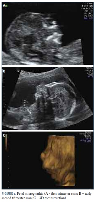
Here we present a case of a healthy primipara woman with a suspicion of fetal micrognathia during the first trimester ultrasound scan. We performed an early second trimester ultrasound scan that confirmed our suspicion of micrognathia and showed it as an isolated finding (see <a href="#f1">Figure 1</a>). The fetal echocardiography was normal and the fetus had normal body movements. Prenatal counselling was made and an invasive diagnostic test was offered and accepted by the couple. The fetal karyotype and array comparative genomic hybridization (CGH) were normal. The couple has requested pregnancy interruption, which has been accepted by the technical certification committee. The anatomic-pathological exam of the fetus showed mandibular hypoplasia, microglossia and a right lung with four lobes. A genetic condition capable of explaining these findings could not been found.</p>     <p>&nbsp;</p>     <p align="center"><a name="f1"></a><img src="/img/revistas/aogp/v9n5/9n5a12f1.jpg"/></p>     
<p>&nbsp;</p>     <p>Fetal micrognathia may be associated with adverse environmental factors, multiple genetic syndromes, such as Pierre Robin sequence, agnathia-microstomia&#8212;synotia syndrome (otocephaly) and the cerebro-costo&#8212;mandibular syndrome and with chromosomal abnormalities, such as trisomy 13 or 18. The incidence of chromosomal abnormalities ranges between 31% and 82% in different studies, but in those cases there are usually other associated fetal anomalies<sup>1,5</sup>.</p>     ]]></body>
<body><![CDATA[<p>The first approach to fetal micrognathia should include search for other anomalies, drug exposure or familial history of syndromes, fetal karyotype and expose the prognosis to parents. In Pierre Robin&#8217;s sequence, both micrognatia and macroglossia cause the tongue to move upwards, preventing the closure of the palate, which results in cleft palate; this diagnosis was excluded by ultrasound in our case. The fetal microglossia found in the anatomic-pathological exam leads us to believe that there has been an abnormal development of the entire area. Objective evaluation of the micrognathia was not necessary in our case due to its severity. The overall prognosis in these cases seems to be poor, with respiratory distress, upper airway obstruction and feeding problems in the neonate<sup>5</sup>. Pregnancy interruption is an option before viability. When continuation of the pregnancy is chosen, serial growth scans with assessment of the amniotic fluid volume and fetal movements should be performed in order to exclude, for example, neuromuscular diseases. Delivery should occur in a reference centre with immediate paediatric assistance for the newborn, as there may be obstruction of the upper airway<sup>5</sup>. Moreover, paediatric anaesthesiologist should be available, because ex utero intrapartum treatment might be an option in severe cases.</p>     <p>This case illustrates the need of a careful observation of the fetal face during each obstetric ultrasound scan; otherwise serious conditions like this can go unnoticed and be diagnosed only after birth, when it might be too late to provide the necessary assistance.</p>     <p>&nbsp;</p>     <p><b>REFERENCES </b></p>     <!-- ref --><p>1. Paladini D. Fetal micrognathia: almost always an ominous finding. Ultrasound Obstet Gynecol. 2010;35(4):377-384.    &nbsp;&nbsp;&nbsp;&nbsp;&nbsp;&nbsp;&nbsp;&nbsp;[&#160;<a href="javascript:void(0);" onclick="javascript: window.open('/scielo.php?script=sci_nlinks&ref=1854000&pid=S1646-5830201500040001200001&lng=','','width=640,height=500,resizable=yes,scrollbars=1,menubar=yes,');">Links</a>&#160;]<!-- end-ref --></p>     <!-- ref --><p>2. Paladini D, Morra T, Teodoro A, Lamberti A, Tremolaterra F, Martinelli P. Objective diagnosis of micrognathia in the fetus: the Jaw Index. Obstet Gynecol 1999;93:382&#8211;386.    &nbsp;&nbsp;&nbsp;&nbsp;&nbsp;&nbsp;&nbsp;&nbsp;[&#160;<a href="javascript:void(0);" onclick="javascript: window.open('/scielo.php?script=sci_nlinks&ref=1854002&pid=S1646-5830201500040001200002&lng=','','width=640,height=500,resizable=yes,scrollbars=1,menubar=yes,');">Links</a>&#160;]<!-- end-ref --></p>     <!-- ref --><p>3. Otto C, Platt LD. The fetal mandible measurement: an objective determination of fetal jaw size. Ultrasound Obstet Gynecol 1991;1:12&#8211;17.    &nbsp;&nbsp;&nbsp;&nbsp;&nbsp;&nbsp;&nbsp;&nbsp;[&#160;<a href="javascript:void(0);" onclick="javascript: window.open('/scielo.php?script=sci_nlinks&ref=1854004&pid=S1646-5830201500040001200003&lng=','','width=640,height=500,resizable=yes,scrollbars=1,menubar=yes,');">Links</a>&#160;]<!-- end-ref --></p>     ]]></body>
<body><![CDATA[<!-- ref --><p>4. Rotten D, Levaillant JM, Martinez H, Ducou H, Le Pointe D, Vicaut E. The fetal mandible: a 2D and 3D sonographic approach to the diagnosis of retrognathia and micrognathia. Ultrasound Obstet Gynecol 2002; 19:122&#8211;130.    &nbsp;&nbsp;&nbsp;&nbsp;&nbsp;&nbsp;&nbsp;&nbsp;[&#160;<a href="javascript:void(0);" onclick="javascript: window.open('/scielo.php?script=sci_nlinks&ref=1854006&pid=S1646-5830201500040001200004&lng=','','width=640,height=500,resizable=yes,scrollbars=1,menubar=yes,');">Links</a>&#160;]<!-- end-ref --></p>     <!-- ref --><p>5. Bromley B, Benacerraf BR. Fetal micrognathia: associated anomalies and outcome. J Ultrasound Med. 1994;13(7):529-533.    &nbsp;&nbsp;&nbsp;&nbsp;&nbsp;&nbsp;&nbsp;&nbsp;[&#160;<a href="javascript:void(0);" onclick="javascript: window.open('/scielo.php?script=sci_nlinks&ref=1854008&pid=S1646-5830201500040001200005&lng=','','width=640,height=500,resizable=yes,scrollbars=1,menubar=yes,');">Links</a>&#160;]<!-- end-ref --></p>     <p>&nbsp;</p>     <p><a href="#topc0">Endere&ccedil;o para correspond&ecirc;ncia</a> | <a href="#topc0">Direcci&oacute;n para correspondencia</a> | <a href="#topc0">Correspondence</a><a name="c0"></a></p>     <p>Ana Galv&#227;o</p>     <p>Centro Materno Infantil do Norte</p>     <p>Centro Hospitalar do Porto</p>     <p>Largo Prof. Abel Salazar</p>     ]]></body>
<body><![CDATA[<p>4099-001 Porto</p>     <p>E-mail: <a href="mailto:ana.m.galvao@gmail.com">ana.m.galvao@gmail.com</a></p>     <p>&nbsp;</p>     <p><b>Recebido em: </b> 01-10-2015<b></b></p>     <p><b>Aceite para publica&#231;&#227;o: </b> 02-11-2015</p>      ]]></body><back>
<ref-list>
<ref id="B1">
<label>1</label><nlm-citation citation-type="journal">
<person-group person-group-type="author">
<name>
<surname><![CDATA[Paladini]]></surname>
<given-names><![CDATA[D]]></given-names>
</name>
</person-group>
<article-title xml:lang="en"><![CDATA[Fetal micrognathia: almost always an ominous finding]]></article-title>
<source><![CDATA[Ultrasound Obstet Gynecol]]></source>
<year>2010</year>
<volume>35</volume>
<numero>4</numero>
<issue>4</issue>
<page-range>377-384</page-range></nlm-citation>
</ref>
<ref id="B2">
<label>2</label><nlm-citation citation-type="journal">
<person-group person-group-type="author">
<name>
<surname><![CDATA[Paladini]]></surname>
<given-names><![CDATA[D]]></given-names>
</name>
<name>
<surname><![CDATA[Morra]]></surname>
<given-names><![CDATA[T]]></given-names>
</name>
<name>
<surname><![CDATA[Teodoro]]></surname>
<given-names><![CDATA[A]]></given-names>
</name>
<name>
<surname><![CDATA[Lamberti]]></surname>
<given-names><![CDATA[A]]></given-names>
</name>
<name>
<surname><![CDATA[Tremolaterra]]></surname>
<given-names><![CDATA[F]]></given-names>
</name>
<name>
<surname><![CDATA[Martinelli]]></surname>
<given-names><![CDATA[P]]></given-names>
</name>
</person-group>
<article-title xml:lang="en"><![CDATA[Objective diagnosis of micrognathia in the fetus: the Jaw Index]]></article-title>
<source><![CDATA[Obstet Gynecol]]></source>
<year>1999</year>
<volume>93</volume>
<page-range>382-386</page-range></nlm-citation>
</ref>
<ref id="B3">
<label>3</label><nlm-citation citation-type="journal">
<person-group person-group-type="author">
<name>
<surname><![CDATA[Otto]]></surname>
<given-names><![CDATA[C]]></given-names>
</name>
<name>
<surname><![CDATA[Platt]]></surname>
<given-names><![CDATA[LD]]></given-names>
</name>
</person-group>
<article-title xml:lang="en"><![CDATA[The fetal mandible measurement: an objective determination of fetal jaw size]]></article-title>
<source><![CDATA[Ultrasound Obstet Gynecol]]></source>
<year>1991</year>
<volume>1</volume>
<page-range>12-17</page-range></nlm-citation>
</ref>
<ref id="B4">
<label>4</label><nlm-citation citation-type="journal">
<person-group person-group-type="author">
<name>
<surname><![CDATA[Rotten]]></surname>
<given-names><![CDATA[D]]></given-names>
</name>
<name>
<surname><![CDATA[Levaillant]]></surname>
<given-names><![CDATA[JM]]></given-names>
</name>
<name>
<surname><![CDATA[Martinez]]></surname>
<given-names><![CDATA[H]]></given-names>
</name>
<name>
<surname><![CDATA[Ducou]]></surname>
<given-names><![CDATA[H]]></given-names>
</name>
<name>
<surname><![CDATA[Le Pointe]]></surname>
<given-names><![CDATA[D]]></given-names>
</name>
<name>
<surname><![CDATA[Vicaut]]></surname>
<given-names><![CDATA[E]]></given-names>
</name>
</person-group>
<article-title xml:lang="en"><![CDATA[The fetal mandible: a 2D and 3D sonographic approach to the diagnosis of retrognathia and micrognathia]]></article-title>
<source><![CDATA[Ultrasound Obstet Gynecol]]></source>
<year>2002</year>
<volume>19</volume>
<page-range>122-130</page-range></nlm-citation>
</ref>
<ref id="B5">
<label>5</label><nlm-citation citation-type="journal">
<person-group person-group-type="author">
<name>
<surname><![CDATA[Bromley]]></surname>
<given-names><![CDATA[B]]></given-names>
</name>
<name>
<surname><![CDATA[Benacerraf]]></surname>
<given-names><![CDATA[BR]]></given-names>
</name>
</person-group>
<article-title xml:lang="en"><![CDATA[Fetal micrognathia: associated anomalies and outcome]]></article-title>
<source><![CDATA[J Ultrasound Med]]></source>
<year>1994</year>
<volume>13</volume>
<numero>7</numero>
<issue>7</issue>
<page-range>529-533</page-range></nlm-citation>
</ref>
</ref-list>
</back>
</article>
