<?xml version="1.0" encoding="ISO-8859-1"?><article xmlns:mml="http://www.w3.org/1998/Math/MathML" xmlns:xlink="http://www.w3.org/1999/xlink" xmlns:xsi="http://www.w3.org/2001/XMLSchema-instance">
<front>
<journal-meta>
<journal-id>1646-5830</journal-id>
<journal-title><![CDATA[Acta Obstétrica e Ginecológica Portuguesa]]></journal-title>
<abbrev-journal-title><![CDATA[Acta Obstet Ginecol Port]]></abbrev-journal-title>
<issn>1646-5830</issn>
<publisher>
<publisher-name><![CDATA[Euromédice, Edições Médicas Lda.]]></publisher-name>
</publisher>
</journal-meta>
<article-meta>
<article-id>S1646-58302017000200009</article-id>
<title-group>
<article-title xml:lang="en"><![CDATA[Myeloid sarcoma involving the cervix: about a clinical case]]></article-title>
<article-title xml:lang="pt"><![CDATA[Sarcoma mielóide do colo do útero: a propósito de um caso clínico]]></article-title>
</title-group>
<contrib-group>
<contrib contrib-type="author">
<name>
<surname><![CDATA[Alves]]></surname>
<given-names><![CDATA[Cláudia Meneses]]></given-names>
</name>
<xref ref-type="aff" rid="A1"/>
</contrib>
<contrib contrib-type="author">
<name>
<surname><![CDATA[Domingues]]></surname>
<given-names><![CDATA[Nelson]]></given-names>
</name>
<xref ref-type="aff" rid="A1"/>
</contrib>
<contrib contrib-type="author">
<name>
<surname><![CDATA[Cruz-Pires]]></surname>
<given-names><![CDATA[Mónica]]></given-names>
</name>
<xref ref-type="aff" rid="A1"/>
</contrib>
<contrib contrib-type="author">
<name>
<surname><![CDATA[Felizardo]]></surname>
<given-names><![CDATA[Diana]]></given-names>
</name>
<xref ref-type="aff" rid="A1"/>
</contrib>
<contrib contrib-type="author">
<name>
<surname><![CDATA[Rosales]]></surname>
<given-names><![CDATA[Maria]]></given-names>
</name>
<xref ref-type="aff" rid="A1"/>
</contrib>
</contrib-group>
<aff id="AA1">
<institution><![CDATA[,Instituto Português de Oncologia do Porto FG, EPE  ]]></institution>
<addr-line><![CDATA[Porto ]]></addr-line>
</aff>
<pub-date pub-type="pub">
<day>00</day>
<month>06</month>
<year>2017</year>
</pub-date>
<pub-date pub-type="epub">
<day>00</day>
<month>06</month>
<year>2017</year>
</pub-date>
<volume>11</volume>
<numero>2</numero>
<fpage>122</fpage>
<lpage>127</lpage>
<copyright-statement/>
<copyright-year/>
<self-uri xlink:href="http://scielo.pt/scielo.php?script=sci_arttext&amp;pid=S1646-58302017000200009&amp;lng=en&amp;nrm=iso"></self-uri><self-uri xlink:href="http://scielo.pt/scielo.php?script=sci_abstract&amp;pid=S1646-58302017000200009&amp;lng=en&amp;nrm=iso"></self-uri><self-uri xlink:href="http://scielo.pt/scielo.php?script=sci_pdf&amp;pid=S1646-58302017000200009&amp;lng=en&amp;nrm=iso"></self-uri><abstract abstract-type="short" xml:lang="en"><p><![CDATA[Myeloid sarcoma is defined as a tumour of myeloblasts and immature myeloid cells that occurs in an extramedullary tissue. It is a rare form of presentation, which may arise in different locations and the cervix is one of the most unusual places. The available therapeutic options are chemotherapy and radiotherapy. The main prognostic factors are the response to initial treatment and cytogenetic abnormalities. This article describes the case of a woman presenting pelvic pain and vaginal discharge as chief complaints. After detection of a mass in the uterine cervix, the diagnosis was established by histology and immunophenotyping]]></p></abstract>
<kwd-group>
<kwd lng="en"><![CDATA[Acute myeloid leukaemia]]></kwd>
<kwd lng="en"><![CDATA[Myeloid sarcoma]]></kwd>
<kwd lng="en"><![CDATA[Cervix]]></kwd>
</kwd-group>
</article-meta>
</front><body><![CDATA[ <p align="right"><font size="2"><b>CASE   REPORT/</B>CASO   CL&#205;NICO</font></p>     <p><font size="4"><b>Myeloid   sarcoma involving the cervix - about a clinical case</b></font></p>     <p><font size="3"><b>Sarcoma   miel&#243;ide do colo do &#250;tero - a prop&#243;sito de um caso cl&#237;nico</b></font></p>     <p><b>Cl&#225;udia   Meneses Alves*, Nelson Domingues**, M&#243;nica Cruz-Pires**, Diana Felizardo*, Maria Rosales**</b></p>     <p>Instituto   Portugu&#234;s de Oncologia do Porto FG, EPE</p>     <p>*Interna de Forma&#231;&#227;o Espec&#237;fica</p>     <p>**Assistente Hospitalar</p>     <p><a href="#c0">Endere&ccedil;o para correspond&ecirc;ncia</a> | <a href="#c0">Direcci&oacute;n para correspondencia</a> | <a href="#c0">Correspondence</a><a name="topc0"></a></p> <hr/>     <p>&nbsp;</p>     <p><b>ABSTRACT</b></p>     ]]></body>
<body><![CDATA[<p>Myeloid   sarcoma is defined as a tumour of myeloblasts and immature myeloid cells that   occurs in an extramedullary tissue. It is a rare form of presentation, which   may arise in different locations and the cervix is one of the most unusual   places. The available therapeutic options are chemotherapy and radiotherapy.   The main prognostic factors are the response to initial treatment and   cytogenetic abnormalities. This article describes the case of a woman   presenting pelvic pain and vaginal discharge as chief complaints. After   detection of a mass in the uterine cervix, the diagnosis was established by   histology and immunophenotyping.</p>     <p><b>Keywords: </b>Acute myeloid   leukaemia; Myeloid sarcoma; Cervix</p> <hr/>     <p>&nbsp;</p>    <p><b>Introduction </b></p>     <p>Acute myeloid leukaemia   (AML) may have different forms of presentation in extramedullary (EM)   locations with or without bone marrow (BM) disease<sup>1</sup>. Myeloid sarcoma   (MS) is a well established form of presentation of AML.&nbsp;It&#180;s defined as a   tumour of myeloblasts or immature myeloid cells that occurs in an EM place<sup>2-13</sup>.   &#8203;It can occur as infiltrates of myeloid blasts in any anatomic site of   the body, however they are only classified as MS if they alter the architecture   of the tissues involved<sup>2,4</sup>.&nbsp;The most common sites of   involvement are subperiosteal bone structures, as well as&nbsp;skin, lymph   nodes, soft tissue, central nervous system, gastrointestinal tract and orbit<sup>2,3,7,10,13-17</sup>.   In the female population, the genital tract and the breasts are sites that may   also be affected, although less frequently<sup>8.14</sup>.&nbsp;MS can be   diagnosed as an isolated form, when there is no previous history of leukaemia,   myelodysplastic or myeloproliferative syndrome and a normal BM aspirate. It   can occur before the onset of AML, concurrently with the diagnosis, or as a   post-treatment stage, as&nbsp;a&nbsp;form of recurrent disease with or without spinal cord involvement<sup>1-7,9,11-13</sup>.</p>     <p>Epidemiologically,   MS has an incidence of 2/1.000.000 in the adult population<sup>7,17</sup>&nbsp;and   is described in approximately 2.5 to 9.1% of patients with AML<sup>1,3,11,14,15</sup>.</p>     <p>The   case presented here intends to illustrate a rare form of MS presentation and   its most relevant defining characteristics, in terms of pathology, epidemiology, management and prognosis. </p>     <p><b>Clinical case</b></p>     <p>Female   patient, 53-year-old, caucasian, with the following past medical history:   erythema nodosum, bilateral episcleritis, disc herniation, depressive   syndrome, pericardial effusion history. Unremarkable social and family   history.&nbsp;She had her menarche at the age of 12, gravida 3, para 2,   abortion 1 and menopause at 49 years old. At the time of diagnosis, she was   taking tibolone as hormonal replacement treatment and was applying topical vaginal oestrogen cream.</p>     <p>Present   illness dates back from April 2015, when she had an episode of vaginal   bleeding. The only anomaly reported then was an intramural pericentimetric   myoma that the pelvic ultrasound exposed.&nbsp;In June 2015 the patient   complained of yellow leucorrhoea associated with increasing pelvic pain.&nbsp;<i>Gardnerella     vaginallis </i>was identified<i> </i>in the exsudate (treated with clindamycin)<i>.</i> Due to persistent complaints, a colpocytology was undertaken in August and the   result was negative for intraepithelial lesion (NILM) with inflammation. The   finding of a hard cervix with easy bleeding led to colposcopy with biopsy of   the cervix and curettage of the cervical canal. The anatomopathological   examination showed MS, triggering the referral to our Institution. The physical   examination was unremarkable, except for the gynaecological examination, that   revealed a yellow leucorrhoea and an enlarged and hardened cervix with 5-6cm of   diameter, although the superficial epithelium was smooth, without erosions or   proliferative lesions. The digital rectal examination detected only an enlarged cervix with apparently spared parametria.
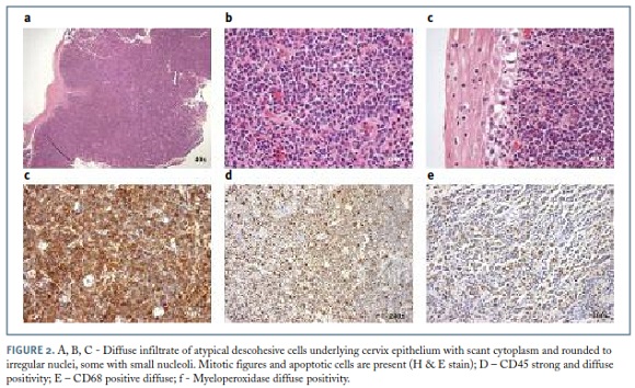
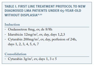
</p>     ]]></body>
<body><![CDATA[<p>The   blood count, serum biochemistry, chest x-ray and electrocardiogram were   normal.&nbsp;The BM aspirate for myelogram and the immunophenotype study by   cytometry revealed no abnormalities. A computed tomography (CT) scan of the   thorax, abdominal and pelvis found &#8220;a tissue density mass on the cervix, with   6.5 cm of transverse length and 4.8 cm of longitudinal length, suggesting a   neoplasic process, coexisting adenopathies with 15x9mm in the right common   iliac chain and in&nbsp;the&nbsp;left external iliac situation with 18 x 12 mm&#8221;   (<a href="#f1">Figure 1</a>). A review of the biopsy slides and a new biopsy of the cervix were   performed and both confirmed the diagnosis. Immunohistochemistry confirmed the result (immunophenotype: CD45 +, CD68 +, MPO +, CD34-, CD20-, CD3-) (<a href="#f2">Figure 2</a>).&nbsp; </p>     <p>&nbsp;</p>    <p align="center"><a name="f1"></a><img src="/img/revistas/aogp/v11n2/11n2a09f1.jpg"/></p>    
<p>&nbsp;</p>    <p align="center"><a name="f2"></a><img src="/img/revistas/aogp/v11n2/11n2a09f2.jpg"/></p>    
<p>&nbsp;</p>     <p>
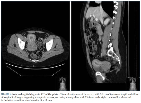
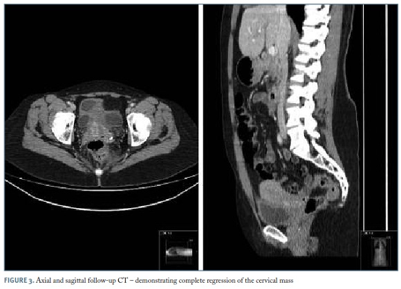
The   treatment was set in a joint meeting between onco-hematology and radio-oncology   units. Initially, the patient started with a chemotherapy &#8220;7+3&#8221; protocol   (idarubicin and cytarabine), followed by consolidation with cytarabine in high   dose according to the Institution protocol for AML (<a href="#t1">Table I</a>) and concluded   with pelvic external radiotherapy&nbsp;for&nbsp;a total of 30Gy.&nbsp;The   hospitalization period for receiving induction chemotherapy and to allow   hematologic recovery was 31 days. The most relevant complications of the   induction chemotherapy were an episode of neutropenic enterocolitis with need   for parenteral nutrition and a severe transfusion reaction, both resolved   without sequelae. The assessment of the response by clinical examination and   CT scan after treatment, showed regression of the cervical and ganglionary   changes observed initially, remaining only a discrete thickening of the cervix   (<a href="#f3">Figure 3</a>). Based on this assessment, we consider that the patient had a   complete response to the treatment.&nbsp;Currently, she is asymptomatic and   maintains follow-up visits in onco-hematology and gynecology units, with a 3   months&#8217; interval between them with focus in blood count and gynaecological examination.</p>     <p>&nbsp;</p>    <p align="center"><a name="t1"></a><img src="/img/revistas/aogp/v11n2/11n2a09t1.jpg"/></p>    
<p>&nbsp;</p>    ]]></body>
<body><![CDATA[<p align="center"><a name="f3"></a><img src="/img/revistas/aogp/v11n2/11n2a09f3.jpg"/></p>    
<p>&nbsp;</p>     <p><b>Discussion</b></p>     <p>The   granulocytic sarcoma (GS), also called chloroma, myeloblastoma, extramedullary   myeloid tumour and monocytic sarcoma, was first described in 1911 by Burns. In   1953, King renamed it chloroma because of its yellowish green appearance,   secondary to high levels of myeloperoxidase.&nbsp;Dock and Warthin recognized   its association with the LMA in 1904.&nbsp;In 1966, Rappaport established the   nomenclature of GS and described it as a destructive solid tumour composed by   immature cells of the granulocytic lineage.&nbsp;Later, in 1988, this   nomenclature had been amended by Davey to extramedullary myeloid tumour   cells.&nbsp;In 2001, the World Health Organization (WHO) set a new nomenclature   for this entity, renaming the myeloid sarcoma and classifying it into two categories: the granulocytic sarcoma and monoblastic sarcoma<sup>6,14</sup>.&nbsp;</p>     <p>According   to the literature,&nbsp;the female genital tract, is an unusual place for MS<sup>7-9,14</sup>.&nbsp;In   this anatomical area, the ovary is the most affected organ (about 36.4%)<sup>7</sup>&nbsp;followed   by the cervix and body of the uterus (about 10%)<sup>14</sup>.&nbsp;</p>     <p>The   clinical presentation of this condition can be diverse and related to the   affected site and the size of the mass. Therefore, the symptoms are caused by   compression with pain, bleeding and, sometimes, systemic symptoms possibly   occurring<sup>1,4,6,14</sup>.&nbsp;In our case, we found local symptoms and   growing abdominal pain but absence of systemic symptoms.</p>     <p>For   the diagnosis,&nbsp;the imaging studies such as CT scan and ultrasound are   useful but not specific and, in most cases, only confirming the presence of a   mass<sup>14</sup>.&nbsp;The diagnosis is made &#8203;&#8203;by histology and by   immunohistochemical and immunophenotyping studies.&nbsp;Morphologically, MS   is characterized by diffuse proliferation of primitive myeloid cells with oval,   irregular, reniform or folded nuclei, fine chromatin, distinct to proeminent   nucleoli and scant to moderate cytoplasm<sup>1,8,20</sup>.&nbsp;The diagnostic   accuracy is aided by immunohistochemical and immunophenotypic studies.   According to the WHO classification of 2008, cytochemical colorations include   chloroacetate esterase, myeloperoxidase and nonspecific esterase.&nbsp;The   immunophenotypic panel may comprise CD68/KP1, MPO, CD117, CD99, CD68/PG-M1,   lysozyme, CD34, TdT, CD56, CD61, CD30, glycophorin and CD4. This study can be   performed in paraffin sections or by fluorescence activated cell sorting   (FACS).&nbsp;The panel must be added with B and T lineage markers for   differential diagnosis<sup>4,5,7</sup>. In the present case, we highlight the   importance of obtaining a definitive diagnosis before any treatment procedures,   in order to apply the most adequate systemic treatment and avoid inappropriate   surgery which could cause disease dissemination<sup>7</sup>.&nbsp;In addition   to these diagnostic elements, literature describes cytogenetic changes   associated with MS in about 50% of the cases.&nbsp;The most frequent changes   are inv(16) and involving chromosome 8 in particular, t(8; 21) (q22; q22),   suggesting that the genomic changes are more frequent than previously   characterized.&nbsp;The prevalence of MS in patients with t(8; 21) is about   9-35%. On the other hand, t(8; 21) is detected in 3.3-43% of patients with MS<sup>4,14</sup>.&nbsp;The cytogenetic study was inconclusive by lack of metaphasis.</p>     <p>The   recommended treatment regimen in patients with isolated MS or concomitant with   AML is the conventional AML chemotherapy protocols, based on evidence of a   higher rate of progression to AML in isolated MS patients receiving localized   treatment (88-100%), when comparing with patients given systemic   chemotherapy (42%)<sup>4</sup>.&nbsp;Although, it is widely established that   systemic treatment used to treat AML patients should be given to isolated MS   patients, the optimal therapeutic option is still unclear<sup>7</sup>. Other   therapeutic approaches such as post-remission treatment with external   radiotherapy, as well as, hematopoietic cell transplantation, are, however, controversial<sup>1,4,7</sup>.&nbsp;</p>     <p>Due   to the fact that MS is a rare disease, it is difficult to establish prognostic   factors<sup>4</sup>.&nbsp;The data on the initial AML remission rate for   patients aged 45-65 years is about 60%<sup>18</sup>. The isolated form of MS,   appears to have&nbsp;a&nbsp;better prognosis than when it arises in association   with AML or as&nbsp;relapse<sup>4</sup>.&nbsp;Cytogenetic abnormalities, such   as t(8; 21), inv(16) and a good response to the initial treatment, both   influence the prognosis positively<sup>14,19</sup>.&nbsp;Delay of the   appropriate treatment has negative impacts on prognosis. If left untreated, on   average, more than 50% of patients will develop AML in a short time period   (5-11 months)<sup>6,7</sup>.&nbsp;In the present case, we expected a favourable   prognosis because it was an isolated form of MS and it showed a favourable   initial response to the therapy.</p>     <p>In   conclusion, with this case report, the authors aim to highlight a rare form of   AML presentation and its diagnostic process, where it was essential the   gynaecological examination together with the laboratory and imaging studies, in   order to make an accurate clinical judgement and define a diagnosis, as well as   the correct treatment to improve the life of this patient.</p>     ]]></body>
<body><![CDATA[<p>&nbsp;</p>     <p>&nbsp; </p>     <p><b>REFERENCES</b></p>     <p>1.   Bakst RL, Tallman MS, Douer D, Yahalom J. How I treat extramedullary acute   myeloid leukaemia.&nbsp;Blood. 2011 Oct; 118, 3785-3794.</p>     <p>2.   Vecchio R,&nbsp;Intagliata E,&nbsp;Fiumara PF,&nbsp;Villari L,&nbsp;Marchese   S,&nbsp;Cacciola E. A rare case of myeloid sarcoma presenting the anal fissure.   G Chir. 2015 Sep-Oct; 36, 222-224.</p>     <!-- ref --><p>3.   Alexiev BA, Wang W, Ning Y, Chumsri S, Gojo I, Rodgers WH, et al. Myeloid   sarcomas:&nbsp;a&nbsp;histologic, immunohistochemical, and cytogenetic   study.&nbsp;Diagnostic Pathology. 2007 Oct;&nbsp;2, 42.    &nbsp;&nbsp;&nbsp;&nbsp;&nbsp;&nbsp;&nbsp;&nbsp;[&#160;<a href="javascript:void(0);" onclick="javascript: window.open('/scielo.php?script=sci_nlinks&ref=1863409&pid=S1646-5830201700020000900003&lng=','','width=640,height=500,resizable=yes,scrollbars=1,menubar=yes,');">Links</a>&#160;]<!-- end-ref --></p>     <!-- ref --><p>4.   Avni B, Koren-Michowitz M. Myeloid sarcoma: current approach and therapeutic   options.&nbsp;Ther&nbsp;Adv Hematol. 2011 Oct;&nbsp;2(5), 309-316.    &nbsp;&nbsp;&nbsp;&nbsp;&nbsp;&nbsp;&nbsp;&nbsp;[&#160;<a href="javascript:void(0);" onclick="javascript: window.open('/scielo.php?script=sci_nlinks&ref=1863411&pid=S1646-5830201700020000900004&lng=','','width=640,height=500,resizable=yes,scrollbars=1,menubar=yes,');">Links</a>&#160;]<!-- end-ref --></p>     <!-- ref --><p>5.   Campidelli C, Agostinelli C, Stitson R, Pileri SA. Myeloid sarcoma:   extramedullary manifestation of myeloid disorders.&nbsp;Am J Clin&nbsp;Pathol.   2009 Sep;&nbsp;132, 426-437.    &nbsp;&nbsp;&nbsp;&nbsp;&nbsp;&nbsp;&nbsp;&nbsp;[&#160;<a href="javascript:void(0);" onclick="javascript: window.open('/scielo.php?script=sci_nlinks&ref=1863413&pid=S1646-5830201700020000900005&lng=','','width=640,height=500,resizable=yes,scrollbars=1,menubar=yes,');">Links</a>&#160;]<!-- end-ref --></p>     <p>6.   Esper RC, S&#225;nchez PE, Bermejo JM. Sarcoma granuloc&#237;tico en una paciente con   leucemia mieloide cr&#243;nica. Revista de la Facultad de Medicina de la UNAM. 2012   Ene-Feb; 55(1),&nbsp;22-28.</p>     <!-- ref --><p>7.   Yu Y,&nbsp;Qin X, Yan S, Wang W, Sun Y, Zhang M. Non-leukemic myeloid sarcoma   Involving the vulva, vagina, and cervix: a case report and literature   review.&nbsp;Onco&nbsp;Targets and Ther. 2015 Dec;&nbsp;8, 3707-3713.    &nbsp;&nbsp;&nbsp;&nbsp;&nbsp;&nbsp;&nbsp;&nbsp;[&#160;<a href="javascript:void(0);" onclick="javascript: window.open('/scielo.php?script=sci_nlinks&ref=1863416&pid=S1646-5830201700020000900007&lng=','','width=640,height=500,resizable=yes,scrollbars=1,menubar=yes,');">Links</a>&#160;]<!-- end-ref --></p>     <!-- ref --><p>8.   Garcia MG, Deavers MT, Knoblock RJ, Chen W, Tsimberidou AM, Manning JT, et   al.&nbsp;Myeloid sarcoma involving the gynecologic - a report of 11 cases   and review of the literature. Am&nbsp;J Clin&nbsp;Pathol 2006 May;&nbsp;125,   783-790.    &nbsp;&nbsp;&nbsp;&nbsp;&nbsp;&nbsp;&nbsp;&nbsp;[&#160;<a href="javascript:void(0);" onclick="javascript: window.open('/scielo.php?script=sci_nlinks&ref=1863418&pid=S1646-5830201700020000900008&lng=','','width=640,height=500,resizable=yes,scrollbars=1,menubar=yes,');">Links</a>&#160;]<!-- end-ref --></p>     <!-- ref --><p>9.   Gill H, Loong F, Mak V, Chan K, Au WY, Kwong YL.&nbsp;Myeloid sarcoma of the   uterine cervix presenting as missed abortion. Arch&nbsp;Gynecol&nbsp;Obstet.   2012 Jul;&nbsp;286, 1339-1341.    &nbsp;&nbsp;&nbsp;&nbsp;&nbsp;&nbsp;&nbsp;&nbsp;[&#160;<a href="javascript:void(0);" onclick="javascript: window.open('/scielo.php?script=sci_nlinks&ref=1863420&pid=S1646-5830201700020000900009&lng=','','width=640,height=500,resizable=yes,scrollbars=1,menubar=yes,');">Links</a>&#160;]<!-- end-ref --></p>     <!-- ref --><p>10.   Ding J, Li H, Qi YK, Wu J, Liu ZB, Huang BC, et al.&nbsp;Ovarian granulocytic   sarcoma of the primary manifestation of acute myelogenous   leukaemia.&nbsp;Int.&nbsp;J Clin&nbsp;Exp Pathol. 2015 Oct;&nbsp;8(10),   13552-13556.    &nbsp;&nbsp;&nbsp;&nbsp;&nbsp;&nbsp;&nbsp;&nbsp;[&#160;<a href="javascript:void(0);" onclick="javascript: window.open('/scielo.php?script=sci_nlinks&ref=1863422&pid=S1646-5830201700020000900010&lng=','','width=640,height=500,resizable=yes,scrollbars=1,menubar=yes,');">Links</a>&#160;]<!-- end-ref --></p>     ]]></body>
<body><![CDATA[<!-- ref --><p>11.   Silva HF,&nbsp;Benevides VM,&nbsp;Ribeiro RM, Franck G;&nbsp;Castro   AJM,&nbsp;Filho FDR,&nbsp;et al. Granulocytic sarcoma with skin and lymph node   initial presentation.&nbsp;Rev Bras Hematol Hemoter. 2008 Feb; 30(1), 61-69.    &nbsp;&nbsp;&nbsp;&nbsp;&nbsp;&nbsp;&nbsp;&nbsp;[&#160;<a href="javascript:void(0);" onclick="javascript: window.open('/scielo.php?script=sci_nlinks&ref=1863424&pid=S1646-5830201700020000900011&lng=','','width=640,height=500,resizable=yes,scrollbars=1,menubar=yes,');">Links</a>&#160;]<!-- end-ref --></p>     <!-- ref --><p>12.   Ruiz MAC, Oca AVM, Rubia MJB, Mart&#237;n AAL.&nbsp;Disfon&#237;a y lumbalgia como   presentaci&#243;n de sarcoma granuloc&#237;tico.&nbsp;Primera manifestaci&#243;n de Leucemia   Mieloide Aguda. Rev Cl&#237;n Med Fam. 2009&nbsp;Jun; 2(7), 368-370.    &nbsp;&nbsp;&nbsp;&nbsp;&nbsp;&nbsp;&nbsp;&nbsp;[&#160;<a href="javascript:void(0);" onclick="javascript: window.open('/scielo.php?script=sci_nlinks&ref=1863426&pid=S1646-5830201700020000900012&lng=','','width=640,height=500,resizable=yes,scrollbars=1,menubar=yes,');">Links</a>&#160;]<!-- end-ref --></p>     <!-- ref --><p>13.   Seo IS, Hull MT,&nbsp;Pak HY. Granulocytic sarcoma of the cervix as a primary   manifestation.&nbsp;Cancer. 1977 Dec;&nbsp;40, 3030-3037.    &nbsp;&nbsp;&nbsp;&nbsp;&nbsp;&nbsp;&nbsp;&nbsp;[&#160;<a href="javascript:void(0);" onclick="javascript: window.open('/scielo.php?script=sci_nlinks&ref=1863428&pid=S1646-5830201700020000900013&lng=','','width=640,height=500,resizable=yes,scrollbars=1,menubar=yes,');">Links</a>&#160;]<!-- end-ref --></p>     <p>14.   Soto GG, Rinc&#243;n JJP, Casta&#241;o JA, Torres JD, G&#243;mez EI. Leucemia mieloide   extramedular con compromiso endometrial: reporte de un caso y revisi&#243;n de la   literatura.&nbsp;Rev&nbsp;Colomb&nbsp;Obstet&nbsp;Gynecol. 2007   Jul-Sep;&nbsp;58 (3), 245-248.</p>     <!-- ref --><p>15.   Hagen PA, Singh C, Hart M, Blaes AH. Differential diagnosis of isolated myeloid   sarcoma: a case report and review of the   Literature.&nbsp;Hematol&nbsp;Rep.&nbsp;2015 Jun;7, 27-30.    &nbsp;&nbsp;&nbsp;&nbsp;&nbsp;&nbsp;&nbsp;&nbsp;[&#160;<a href="javascript:void(0);" onclick="javascript: window.open('/scielo.php?script=sci_nlinks&ref=1863431&pid=S1646-5830201700020000900015&lng=','','width=640,height=500,resizable=yes,scrollbars=1,menubar=yes,');">Links</a>&#160;]<!-- end-ref --></p>     <!-- ref --><p>16.   Aguiar TGS, Oliveira IFL, Guimar&#227;es CVN, Adad, SJ, Moraes-Souza H. Sarcoma   granuloc&#237;tico multic&#234;ntrico como recidiva de leucemia mieloide aguda.&nbsp;Rev   bras &nbsp;Hematol&nbsp;Hemoter. 2009 Jun;&nbsp;31, 207-210.    &nbsp;&nbsp;&nbsp;&nbsp;&nbsp;&nbsp;&nbsp;&nbsp;[&#160;<a href="javascript:void(0);" onclick="javascript: window.open('/scielo.php?script=sci_nlinks&ref=1863433&pid=S1646-5830201700020000900016&lng=','','width=640,height=500,resizable=yes,scrollbars=1,menubar=yes,');">Links</a>&#160;]<!-- end-ref --></p>     <!-- ref --><p>17.   Wang H, Wang H, Ma Q, Chen Y. Intracranial granulocytic sarcoma: two cases and   literature review.&nbsp;Int J Clin Exp Med. 2015 Oct; 8(10), 19586-19590.    &nbsp;&nbsp;&nbsp;&nbsp;&nbsp;&nbsp;&nbsp;&nbsp;[&#160;<a href="javascript:void(0);" onclick="javascript: window.open('/scielo.php?script=sci_nlinks&ref=1863435&pid=S1646-5830201700020000900017&lng=','','width=640,height=500,resizable=yes,scrollbars=1,menubar=yes,');">Links</a>&#160;]<!-- end-ref --></p>     <!-- ref --><p>18.   Hoffbrand AV, Moss PAH. Fundamentos em Hematologia. 6&#170; edi&#231;&#227;o. Artmed. 2013.    &nbsp;&nbsp;&nbsp;&nbsp;&nbsp;&nbsp;&nbsp;&nbsp;[&#160;<a href="javascript:void(0);" onclick="javascript: window.open('/scielo.php?script=sci_nlinks&ref=1863437&pid=S1646-5830201700020000900018&lng=','','width=640,height=500,resizable=yes,scrollbars=1,menubar=yes,');">Links</a>&#160;]<!-- end-ref --></p>     <!-- ref --><p>19.   Lichtman MA, Kaushansky K, Kipps TJ, Prchal JT, Levi MM.&nbsp;Williams Manual   of Hematology, 8th edition. McGraw-Hill. 2010.    &nbsp;&nbsp;&nbsp;&nbsp;&nbsp;&nbsp;&nbsp;&nbsp;[&#160;<a href="javascript:void(0);" onclick="javascript: window.open('/scielo.php?script=sci_nlinks&ref=1863439&pid=S1646-5830201700020000900019&lng=','','width=640,height=500,resizable=yes,scrollbars=1,menubar=yes,');">Links</a>&#160;]<!-- end-ref --></p>     <!-- ref --><p>20.   Kurman RJ, Carcangiu ML, Herrington CS, Young RH. WHO Classification of Tumours   of Female Reproductive Organs. 4th edition. WHO. 2014.    &nbsp;&nbsp;&nbsp;&nbsp;&nbsp;&nbsp;&nbsp;&nbsp;[&#160;<a href="javascript:void(0);" onclick="javascript: window.open('/scielo.php?script=sci_nlinks&ref=1863441&pid=S1646-5830201700020000900020&lng=','','width=640,height=500,resizable=yes,scrollbars=1,menubar=yes,');">Links</a>&#160;]<!-- end-ref --></p>     <!-- ref --><p>21.   Mayer RJ, Davis RB, Schiffer CA, Berg DT, Powell BL, Schulman P, et al.   Intensive Postremission Chemotherapy in Adults with Acute Myeloid Leukaemia for   The Cancer and Leukemia Group B. N Engl J M. 1994; 331, 896-903.    &nbsp;&nbsp;&nbsp;&nbsp;&nbsp;&nbsp;&nbsp;&nbsp;[&#160;<a href="javascript:void(0);" onclick="javascript: window.open('/scielo.php?script=sci_nlinks&ref=1863443&pid=S1646-5830201700020000900021&lng=','','width=640,height=500,resizable=yes,scrollbars=1,menubar=yes,');">Links</a>&#160;]<!-- end-ref --></p>     <p>22.   Wiernik PH, Banks PLC, Case Jr DC, Arlin ZA, Periman PO, Todd MB, et al.   Cytarabine Plus Idarubicin or Daunorubicin as Induction and Consolidation   Therapy for Previously Untreated Adult Patients With Acute Myeloid Leukaemia.   Blood. 1992 January; 79, No 2, 313-319. </p>     <p>&nbsp;</p>     <p><a href="#topc0">Endere&ccedil;o para correspond&ecirc;ncia</a> | <a href="#topc0">Direcci&oacute;n para correspondencia</a> | <a href="#topc0">Correspondence</a><a name="c0"></a></p>     <p>Cl&#225;udia   Meneses Alves</p>     <p>E-mail: <a href="mailto:menesesclaudia@gmail.com">menesesclaudia@gmail.com</a></p>     <p>&nbsp;</p>     <p><b>Acknowledgements</b></p>     <p>The   authors would like to thank Dra. Fernanda Caiano (gynaecologist), the first   doctor to examine the patient and suspect the diagnostic, Dra. Isabel Macedo   Pinto (pathologist) who, firstly, defined the anathomopathological diagnostic   and Dra. Susana F&#233;lix (radiologist) for advising on some of the CT scan   figures. </p>     <p>&nbsp;</p>     ]]></body>
<body><![CDATA[<p>Recebido   em: 12/05/2016</p>     <p>Aceite para   publica&#231;&#227;o: 06/01/2017</p>      ]]></body><back>
<ref-list>
<ref id="B1">
<label>1</label><nlm-citation citation-type="journal">
<person-group person-group-type="author">
<name>
<surname><![CDATA[Bakst]]></surname>
<given-names><![CDATA[RL]]></given-names>
</name>
<name>
<surname><![CDATA[Tallman]]></surname>
<given-names><![CDATA[MS]]></given-names>
</name>
<name>
<surname><![CDATA[Douer]]></surname>
<given-names><![CDATA[D]]></given-names>
</name>
<name>
<surname><![CDATA[Yahalom]]></surname>
<given-names><![CDATA[J]]></given-names>
</name>
</person-group>
<article-title xml:lang="en"><![CDATA[How I treat extramedullary acute myeloid leukaemia]]></article-title>
<source><![CDATA[Blood]]></source>
<year>2011</year>
<volume>118</volume>
<page-range>3785-3794</page-range></nlm-citation>
</ref>
<ref id="B2">
<label>2</label><nlm-citation citation-type="journal">
<person-group person-group-type="author">
<name>
<surname><![CDATA[Vecchio]]></surname>
<given-names><![CDATA[R]]></given-names>
</name>
<name>
<surname><![CDATA[Intagliata]]></surname>
<given-names><![CDATA[E]]></given-names>
</name>
<name>
<surname><![CDATA[Fiumara]]></surname>
<given-names><![CDATA[PF]]></given-names>
</name>
<name>
<surname><![CDATA[Villari]]></surname>
<given-names><![CDATA[L]]></given-names>
</name>
<name>
<surname><![CDATA[Marchese]]></surname>
<given-names><![CDATA[S]]></given-names>
</name>
<name>
<surname><![CDATA[Cacciola]]></surname>
<given-names><![CDATA[E]]></given-names>
</name>
</person-group>
<article-title xml:lang="en"><![CDATA[A rare case of myeloid sarcoma presenting the anal fissure]]></article-title>
<source><![CDATA[G Chir]]></source>
<year>2015</year>
<volume>36</volume>
<page-range>222-224</page-range></nlm-citation>
</ref>
<ref id="B3">
<label>3</label><nlm-citation citation-type="journal">
<person-group person-group-type="author">
<name>
<surname><![CDATA[Alexiev]]></surname>
<given-names><![CDATA[BA]]></given-names>
</name>
<name>
<surname><![CDATA[Wang]]></surname>
<given-names><![CDATA[W]]></given-names>
</name>
<name>
<surname><![CDATA[Ning]]></surname>
<given-names><![CDATA[Y]]></given-names>
</name>
<name>
<surname><![CDATA[Chumsri]]></surname>
<given-names><![CDATA[S]]></given-names>
</name>
<name>
<surname><![CDATA[Gojo]]></surname>
<given-names><![CDATA[I]]></given-names>
</name>
<name>
<surname><![CDATA[Rodgers]]></surname>
<given-names><![CDATA[WH]]></given-names>
</name>
</person-group>
<article-title xml:lang="en"><![CDATA[Myeloid sarcomas: a histologic, immunohistochemical, and cytogenetic study]]></article-title>
<source><![CDATA[Diagnostic Pathology]]></source>
<year>2007</year>
<month>10</month>
<volume>2</volume>
<page-range>42</page-range></nlm-citation>
</ref>
<ref id="B4">
<label>4</label><nlm-citation citation-type="journal">
<person-group person-group-type="author">
<name>
<surname><![CDATA[Avni]]></surname>
<given-names><![CDATA[B]]></given-names>
</name>
<name>
<surname><![CDATA[Koren-Michowitz]]></surname>
<given-names><![CDATA[M]]></given-names>
</name>
</person-group>
<article-title xml:lang="en"><![CDATA[Myeloid sarcoma: current approach and therapeutic options]]></article-title>
<source><![CDATA[Ther Adv Hematol]]></source>
<year>2011</year>
<month>10</month>
<volume>2</volume>
<numero>5</numero>
<issue>5</issue>
<page-range>309-316</page-range></nlm-citation>
</ref>
<ref id="B5">
<label>5</label><nlm-citation citation-type="journal">
<person-group person-group-type="author">
<name>
<surname><![CDATA[Campidelli]]></surname>
<given-names><![CDATA[C]]></given-names>
</name>
<name>
<surname><![CDATA[Agostinelli]]></surname>
<given-names><![CDATA[C]]></given-names>
</name>
<name>
<surname><![CDATA[Stitson]]></surname>
<given-names><![CDATA[R]]></given-names>
</name>
<name>
<surname><![CDATA[Pileri]]></surname>
<given-names><![CDATA[SA]]></given-names>
</name>
</person-group>
<article-title xml:lang="en"><![CDATA[Myeloid sarcoma: extramedullary manifestation of myeloid disorders]]></article-title>
<source><![CDATA[Am J Clin Pathol]]></source>
<year>2009</year>
<month>09</month>
<volume>132</volume>
<page-range>426-437</page-range></nlm-citation>
</ref>
<ref id="B6">
<label>6</label><nlm-citation citation-type="journal">
<person-group person-group-type="author">
<name>
<surname><![CDATA[Esper]]></surname>
<given-names><![CDATA[RC]]></given-names>
</name>
<name>
<surname><![CDATA[Sánchez]]></surname>
<given-names><![CDATA[PE]]></given-names>
</name>
<name>
<surname><![CDATA[Bermejo]]></surname>
<given-names><![CDATA[JM]]></given-names>
</name>
</person-group>
<article-title xml:lang="es"><![CDATA[Sarcoma granulocítico en una paciente con leucemia mieloide crónica]]></article-title>
<source><![CDATA[Revista de la Facultad de Medicina de la UNAM]]></source>
<year>2012</year>
<month>02</month>
<volume>55</volume>
<numero>1</numero>
<issue>1</issue>
<page-range>22-28</page-range></nlm-citation>
</ref>
<ref id="B7">
<label>7</label><nlm-citation citation-type="journal">
<person-group person-group-type="author">
<name>
<surname><![CDATA[Yu]]></surname>
<given-names><![CDATA[Y]]></given-names>
</name>
<name>
<surname><![CDATA[Qin]]></surname>
<given-names><![CDATA[X]]></given-names>
</name>
<name>
<surname><![CDATA[Yan]]></surname>
<given-names><![CDATA[S]]></given-names>
</name>
<name>
<surname><![CDATA[Wang]]></surname>
<given-names><![CDATA[W]]></given-names>
</name>
<name>
<surname><![CDATA[Sun]]></surname>
<given-names><![CDATA[Y]]></given-names>
</name>
<name>
<surname><![CDATA[Zhang]]></surname>
<given-names><![CDATA[M]]></given-names>
</name>
</person-group>
<article-title xml:lang="en"><![CDATA[Non-leukemic myeloid sarcoma Involving the vulva, vagina, and cervix: a case report and literature review]]></article-title>
<source><![CDATA[Onco Targets and Ther]]></source>
<year>2015</year>
<month>12</month>
<volume>8</volume>
<page-range>3707-3713</page-range></nlm-citation>
</ref>
<ref id="B8">
<label>8</label><nlm-citation citation-type="journal">
<person-group person-group-type="author">
<name>
<surname><![CDATA[Garcia]]></surname>
<given-names><![CDATA[MG]]></given-names>
</name>
<name>
<surname><![CDATA[Deavers]]></surname>
<given-names><![CDATA[MT]]></given-names>
</name>
<name>
<surname><![CDATA[Knoblock]]></surname>
<given-names><![CDATA[RJ]]></given-names>
</name>
<name>
<surname><![CDATA[Chen]]></surname>
<given-names><![CDATA[W]]></given-names>
</name>
<name>
<surname><![CDATA[Tsimberidou]]></surname>
<given-names><![CDATA[AM]]></given-names>
</name>
<name>
<surname><![CDATA[Manning]]></surname>
<given-names><![CDATA[JT]]></given-names>
</name>
</person-group>
<article-title xml:lang="en"><![CDATA[Myeloid sarcoma involving the gynecologic - a report of 11 cases and review of the literature]]></article-title>
<source><![CDATA[Am J Clin Pathol]]></source>
<year>2006</year>
<month>05</month>
<volume>125</volume>
<page-range>783-790</page-range></nlm-citation>
</ref>
<ref id="B9">
<label>9</label><nlm-citation citation-type="journal">
<person-group person-group-type="author">
<name>
<surname><![CDATA[Gill]]></surname>
<given-names><![CDATA[H]]></given-names>
</name>
<name>
<surname><![CDATA[Loong]]></surname>
<given-names><![CDATA[F]]></given-names>
</name>
<name>
<surname><![CDATA[Mak]]></surname>
<given-names><![CDATA[V]]></given-names>
</name>
<name>
<surname><![CDATA[Chan]]></surname>
<given-names><![CDATA[K]]></given-names>
</name>
<name>
<surname><![CDATA[Au]]></surname>
<given-names><![CDATA[WY]]></given-names>
</name>
<name>
<surname><![CDATA[Kwong]]></surname>
<given-names><![CDATA[YL]]></given-names>
</name>
</person-group>
<article-title xml:lang="en"><![CDATA[Myeloid sarcoma of the uterine cervix presenting as missed abortion]]></article-title>
<source><![CDATA[Arch Gynecol Obstet]]></source>
<year>2012</year>
<month>07</month>
<volume>286</volume>
<page-range>1339-1341</page-range></nlm-citation>
</ref>
<ref id="B10">
<label>10</label><nlm-citation citation-type="journal">
<person-group person-group-type="author">
<name>
<surname><![CDATA[Ding]]></surname>
<given-names><![CDATA[J]]></given-names>
</name>
<name>
<surname><![CDATA[Li]]></surname>
<given-names><![CDATA[H]]></given-names>
</name>
<name>
<surname><![CDATA[Qi]]></surname>
<given-names><![CDATA[YK]]></given-names>
</name>
<name>
<surname><![CDATA[Wu]]></surname>
<given-names><![CDATA[J]]></given-names>
</name>
<name>
<surname><![CDATA[Liu]]></surname>
<given-names><![CDATA[ZB]]></given-names>
</name>
<name>
<surname><![CDATA[Huang]]></surname>
<given-names><![CDATA[BC]]></given-names>
</name>
</person-group>
<article-title xml:lang="en"><![CDATA[Ovarian granulocytic sarcoma of the primary manifestation of acute myelogenous leukaemia: Int]]></article-title>
<source><![CDATA[J Clin Exp Pathol]]></source>
<year>2015</year>
<month>10</month>
<volume>8</volume>
<numero>10</numero>
<issue>10</issue>
<page-range>13552-13556</page-range></nlm-citation>
</ref>
<ref id="B11">
<label>11</label><nlm-citation citation-type="journal">
<person-group person-group-type="author">
<name>
<surname><![CDATA[Silva]]></surname>
<given-names><![CDATA[HF]]></given-names>
</name>
<name>
<surname><![CDATA[Benevides]]></surname>
<given-names><![CDATA[VM]]></given-names>
</name>
<name>
<surname><![CDATA[Ribeiro]]></surname>
<given-names><![CDATA[RM]]></given-names>
</name>
<name>
<surname><![CDATA[Franck]]></surname>
<given-names><![CDATA[G]]></given-names>
</name>
<name>
<surname><![CDATA[Castro]]></surname>
<given-names><![CDATA[AJM]]></given-names>
</name>
<name>
<surname><![CDATA[Filho]]></surname>
<given-names><![CDATA[FDR]]></given-names>
</name>
</person-group>
<article-title xml:lang="en"><![CDATA[Granulocytic sarcoma with skin and lymph node initial presentation]]></article-title>
<source><![CDATA[Rev Bras Hematol Hemoter]]></source>
<year>2008</year>
<month>02</month>
<volume>30</volume>
<numero>1</numero>
<issue>1</issue>
<page-range>61-69</page-range></nlm-citation>
</ref>
<ref id="B12">
<label>12</label><nlm-citation citation-type="journal">
<person-group person-group-type="author">
<name>
<surname><![CDATA[Ruiz]]></surname>
<given-names><![CDATA[MAC]]></given-names>
</name>
<name>
<surname><![CDATA[Oca]]></surname>
<given-names><![CDATA[AVM]]></given-names>
</name>
<name>
<surname><![CDATA[Rubia]]></surname>
<given-names><![CDATA[MJB]]></given-names>
</name>
<name>
<surname><![CDATA[Martín]]></surname>
<given-names><![CDATA[AAL]]></given-names>
</name>
</person-group>
<article-title xml:lang="es"><![CDATA[Disfonía y lumbalgia como presentación de sarcoma granulocítico: Primera manifestación de Leucemia Mieloide Aguda]]></article-title>
<source><![CDATA[Rev Clín Med Fam]]></source>
<year>2009</year>
<month>06</month>
<volume>2</volume>
<numero>7</numero>
<issue>7</issue>
<page-range>368-370</page-range></nlm-citation>
</ref>
<ref id="B13">
<label>13</label><nlm-citation citation-type="journal">
<person-group person-group-type="author">
<name>
<surname><![CDATA[Seo]]></surname>
<given-names><![CDATA[IS]]></given-names>
</name>
<name>
<surname><![CDATA[Hull]]></surname>
<given-names><![CDATA[MT]]></given-names>
</name>
<name>
<surname><![CDATA[Pak]]></surname>
<given-names><![CDATA[HY]]></given-names>
</name>
</person-group>
<article-title xml:lang="en"><![CDATA[Granulocytic sarcoma of the cervix as a primary manifestation]]></article-title>
<source><![CDATA[Cancer]]></source>
<year>1977</year>
<month>12</month>
<volume>40</volume>
<page-range>3030-3037</page-range></nlm-citation>
</ref>
<ref id="B14">
<label>14</label><nlm-citation citation-type="journal">
<person-group person-group-type="author">
<name>
<surname><![CDATA[Soto]]></surname>
<given-names><![CDATA[GG]]></given-names>
</name>
<name>
<surname><![CDATA[Rincón]]></surname>
<given-names><![CDATA[JJP]]></given-names>
</name>
<name>
<surname><![CDATA[Castaño]]></surname>
<given-names><![CDATA[JA]]></given-names>
</name>
<name>
<surname><![CDATA[Torres]]></surname>
<given-names><![CDATA[JD]]></given-names>
</name>
<name>
<surname><![CDATA[Gómez]]></surname>
<given-names><![CDATA[EI]]></given-names>
</name>
</person-group>
<article-title xml:lang="es"><![CDATA[Leucemia mieloide extramedular con compromiso endometrial: reporte de un caso y revisión de la literatura]]></article-title>
<source><![CDATA[Rev Colomb Obstet Gynecol]]></source>
<year>2007</year>
<month>07</month>
<volume>58</volume>
<numero>3</numero>
<issue>3</issue>
<page-range>245-248</page-range></nlm-citation>
</ref>
<ref id="B15">
<label>15</label><nlm-citation citation-type="journal">
<person-group person-group-type="author">
<name>
<surname><![CDATA[Hagen]]></surname>
<given-names><![CDATA[PA]]></given-names>
</name>
<name>
<surname><![CDATA[Singh]]></surname>
<given-names><![CDATA[C]]></given-names>
</name>
<name>
<surname><![CDATA[Hart]]></surname>
<given-names><![CDATA[M]]></given-names>
</name>
<name>
<surname><![CDATA[Blaes]]></surname>
<given-names><![CDATA[AH]]></given-names>
</name>
</person-group>
<article-title xml:lang="en"><![CDATA[Differential diagnosis of isolated myeloid sarcoma: a case report and review of the Literature]]></article-title>
<source><![CDATA[Hematol Rep]]></source>
<year>2015</year>
<month>06</month>
<volume>7</volume>
<page-range>27-30</page-range></nlm-citation>
</ref>
<ref id="B16">
<label>16</label><nlm-citation citation-type="journal">
<person-group person-group-type="author">
<name>
<surname><![CDATA[Aguiar]]></surname>
<given-names><![CDATA[TGS]]></given-names>
</name>
<name>
<surname><![CDATA[Oliveira]]></surname>
<given-names><![CDATA[IFL]]></given-names>
</name>
<name>
<surname><![CDATA[Guimarães]]></surname>
<given-names><![CDATA[CVN]]></given-names>
</name>
<name>
<surname><![CDATA[Adad]]></surname>
</name>
<name>
<surname><![CDATA[SJ]]></surname>
</name>
<name>
<surname><![CDATA[Moraes-Souza]]></surname>
<given-names><![CDATA[H]]></given-names>
</name>
</person-group>
<article-title xml:lang="pt"><![CDATA[Sarcoma granulocítico multicêntrico como recidiva de leucemia mieloide aguda]]></article-title>
<source><![CDATA[Rev bras Hematol Hemoter]]></source>
<year>2009</year>
<month>06</month>
<volume>31</volume>
<page-range>207-210</page-range></nlm-citation>
</ref>
<ref id="B17">
<label>17</label><nlm-citation citation-type="journal">
<person-group person-group-type="author">
<name>
<surname><![CDATA[Wang]]></surname>
<given-names><![CDATA[H]]></given-names>
</name>
<name>
<surname><![CDATA[Wang]]></surname>
<given-names><![CDATA[H]]></given-names>
</name>
<name>
<surname><![CDATA[Ma]]></surname>
<given-names><![CDATA[Q]]></given-names>
</name>
<name>
<surname><![CDATA[Chen]]></surname>
<given-names><![CDATA[Y]]></given-names>
</name>
</person-group>
<article-title xml:lang="en"><![CDATA[Intracranial granulocytic sarcoma: two cases and literature review]]></article-title>
<source><![CDATA[Int J Clin Exp Med]]></source>
<year>2015</year>
<month>10</month>
<volume>8</volume>
<numero>10</numero>
<issue>10</issue>
<page-range>19586-19590</page-range></nlm-citation>
</ref>
<ref id="B18">
<label>18</label><nlm-citation citation-type="book">
<person-group person-group-type="author">
<name>
<surname><![CDATA[Hoffbrand]]></surname>
<given-names><![CDATA[AV]]></given-names>
</name>
<name>
<surname><![CDATA[Moss]]></surname>
<given-names><![CDATA[PAH]]></given-names>
</name>
</person-group>
<source><![CDATA[Fundamentos em Hematologia]]></source>
<year>2013</year>
<edition>6</edition>
<publisher-name><![CDATA[Artmed]]></publisher-name>
</nlm-citation>
</ref>
<ref id="B19">
<label>19</label><nlm-citation citation-type="book">
<person-group person-group-type="author">
<name>
<surname><![CDATA[Lichtman]]></surname>
<given-names><![CDATA[MA]]></given-names>
</name>
<name>
<surname><![CDATA[Kaushansky]]></surname>
<given-names><![CDATA[K]]></given-names>
</name>
<name>
<surname><![CDATA[Kipps]]></surname>
<given-names><![CDATA[TJ]]></given-names>
</name>
<name>
<surname><![CDATA[Prchal]]></surname>
<given-names><![CDATA[JT]]></given-names>
</name>
<name>
<surname><![CDATA[Levi]]></surname>
<given-names><![CDATA[MM]]></given-names>
</name>
</person-group>
<source><![CDATA[Williams Manual of Hematology]]></source>
<year>2010</year>
<edition>8</edition>
<publisher-name><![CDATA[McGraw-Hill]]></publisher-name>
</nlm-citation>
</ref>
<ref id="B20">
<label>20</label><nlm-citation citation-type="book">
<person-group person-group-type="author">
<name>
<surname><![CDATA[Kurman]]></surname>
<given-names><![CDATA[RJ]]></given-names>
</name>
<name>
<surname><![CDATA[Carcangiu]]></surname>
<given-names><![CDATA[ML]]></given-names>
</name>
<name>
<surname><![CDATA[Herrington]]></surname>
<given-names><![CDATA[CS]]></given-names>
</name>
<name>
<surname><![CDATA[Young]]></surname>
<given-names><![CDATA[RH]]></given-names>
</name>
</person-group>
<source><![CDATA[WHO Classification of Tumours of Female Reproductive Organs]]></source>
<year>2014</year>
<edition>4</edition>
<publisher-name><![CDATA[WHO]]></publisher-name>
</nlm-citation>
</ref>
<ref id="B21">
<label>21</label><nlm-citation citation-type="journal">
<person-group person-group-type="author">
<name>
<surname><![CDATA[Mayer]]></surname>
<given-names><![CDATA[RJ]]></given-names>
</name>
<name>
<surname><![CDATA[Davis]]></surname>
<given-names><![CDATA[RB]]></given-names>
</name>
<name>
<surname><![CDATA[Schiffer]]></surname>
<given-names><![CDATA[CA]]></given-names>
</name>
<name>
<surname><![CDATA[Berg]]></surname>
<given-names><![CDATA[DT]]></given-names>
</name>
<name>
<surname><![CDATA[Powell]]></surname>
<given-names><![CDATA[BL]]></given-names>
</name>
<name>
<surname><![CDATA[Schulman]]></surname>
<given-names><![CDATA[P]]></given-names>
</name>
</person-group>
<article-title xml:lang="en"><![CDATA[Intensive Postremission Chemotherapy in Adults with Acute Myeloid Leukaemia for The Cancer and Leukemia Group B]]></article-title>
<source><![CDATA[N Engl J M]]></source>
<year>1994</year>
<volume>331</volume>
<page-range>896-903</page-range></nlm-citation>
</ref>
<ref id="B22">
<label>22</label><nlm-citation citation-type="journal">
<person-group person-group-type="author">
<name>
<surname><![CDATA[Wiernik]]></surname>
<given-names><![CDATA[PH]]></given-names>
</name>
<name>
<surname><![CDATA[Banks]]></surname>
<given-names><![CDATA[PLC]]></given-names>
</name>
<name>
<surname><![CDATA[Case]]></surname>
<given-names><![CDATA[DC]]></given-names>
</name>
<name>
<surname><![CDATA[Arlin]]></surname>
<given-names><![CDATA[ZA]]></given-names>
</name>
<name>
<surname><![CDATA[Periman]]></surname>
<given-names><![CDATA[PO]]></given-names>
</name>
<name>
<surname><![CDATA[Todd]]></surname>
<given-names><![CDATA[MB]]></given-names>
</name>
</person-group>
<article-title xml:lang="en"><![CDATA[Cytarabine Plus Idarubicin or Daunorubicin as Induction and Consolidation Therapy for Previously Untreated Adult Patients With Acute Myeloid Leukaemia]]></article-title>
<source><![CDATA[Blood]]></source>
<year>1992</year>
<month> J</month>
<day>an</day>
<volume>79</volume>
<numero>2</numero>
<issue>2</issue>
<page-range>313-319</page-range></nlm-citation>
</ref>
</ref-list>
</back>
</article>
