A Rare Cause of Acute Liver Failure: Diffuse Liver Metastization of Merkel Cell Carcinoma
Uma causa rara de falência hepática aguda: metastização difusa de carcinoma de células de Merkel
Ana Luísa Santosa–c, Vítor Magno Pereirad, Rosa Coelhoa–c, Rui Gaspara–c, Hélder Cardosoa–c, Joanne Lopesb,e, Guilherme Macedoa–c
aGastroenterology Department, Centro Hospitalar de São João, Porto, Portugal; bPorto Medical School, University of Porto, Porto, Portugal; cWorld Gastroenterology Organisation (WGO) Oporto Training Center, Porto, Portugal; dGastroenterology Department, Hospital Central do Funchal, Madeira, Portugal; ePathology Department, Centro Hospitalar de São João, Porto, Portugal
* Corresponding author.
ABSTRACT
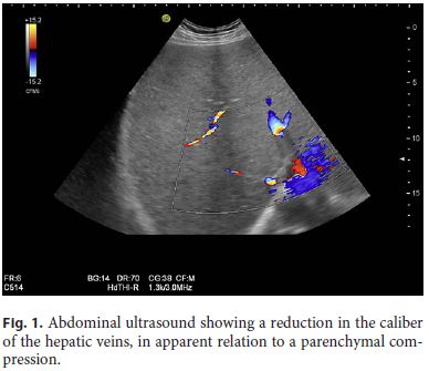
]]> The authors describe a case of a 76-year-old male with a medical history of Merkel cell carcinoma (MCC) of the right lower eyelid. He was admitted to the emergency department due to abdominal pain in the right hypochondrium, nauseas, asthenia, and choluria with 3 days of evolution. Biochemical liver workup revealed a cytocholestase pattern as well as a prolonged prothrombin time. After admission, the patient developed hepatic encephalopathy, and a clinical and analytical worsening was observed. Abdominal ultrasound showed a reduction in the caliber of the hepatic veins, in apparent relation to a parenchymal compression. Liver biopsy was performed and showed an extensive infiltration of the hepatic parenchyma by a solid neoplasm, which, upon immunohistochemical study, was compatible with a diffuse metastization of a MCC. We report this clinical case due to its rarity of presentation and to show the important role of liver biopsy in cases of acute hepatitis.Keywords: Neuroendocrine carcinoma, Liver metástases, Liver biopsy, Liver failure, Hepatic encephalopathy
RESUMO
Os autores descrevem o caso de um homem de 76 anos, com carcinoma de células de Merkel (CCM) da pálpebra direita, admitido no Serviço de Urgência por dor abdominal no hipocôndrio direito, náuseas, astenia e colúria com 3 dias de evolução. Analiticamente, apresentava um padrão citocolestático e coagulopatia com prolongamento do tempo de protrombina. Durante o internamento, desenvolveu encefalopatia hepática, com agravamento clínico e analítico. Ecograficamente, verificou-se redução do calibre das veias heapticas, em aparente relação com compressão parenquimatosa. Foi realizada biópsia hepática que relevou infiltração extensa do parênquima hepático por uma neoplasia sólida compatível, imunohistoquimicamente, com metastização pelo CCM. Os autores reportam este caso clínico devido à raridade da sua apresentação, bem como para salientar o papel da biópsia hepática na abordagem dos casos de hepatite aguda.
Palavras-Chave: Carcinoma neuroendócrino, Metastização hepática, Biópsia hepática, Falência hepática, Encefalopatia hepatica
Introduction
Merkel cell carcinoma (MCC) is a rare tumor of the skin of a neuroendocrine origin. Its estimated annual incidence rate is 0.7 per 100,000 persons, being higher in males, elderly subjects, and people with light skin and those with increased sun exposure [1]. Immunosuppressed patients have also a higher risk of MCC. It presents with a rapidly growing asymptomatic solitary, firm, non-tender, flesh-colored to red tumor with nodular or plaque features. Typically, the diagnosis is made based on incisional tumor biopsy. The main sites of MCC are the head and neck (53%) and extremities (34–35%) [2]. MCC has a high tendency to local recurrence and regional lymph node disease. Distant dissemination occurs in 5–8% of the cases, mainly in the liver and lung and typically as a solitary focus. The sentinel node biopsy is useful to identify patients with occult metastasis [3].
The authors describe a rare case of a MCC with a diffuse liver metastization diagnosed by liver biopsy.
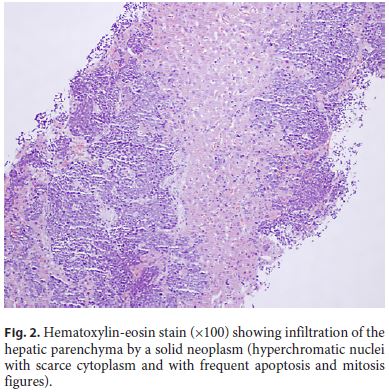
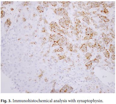
]]> Clinical CaseThe authors describe the case of a 76-year-old male with a medical history of MCC of the right lower eyelid staged pT2NxR1 who underwent surgical treatment. His surgical wound was treated with amoxicillin-clavulanate. Seven days later, the patient was referred to the Emergency Department due to abdominal pain, mainly in the right hypochondrium, jaundice, choluria, nauseas, and asthenia with 3 days of evolution. Analytically, he presented an aspartate aminotransferase of 333 U/L (9 times the upper limit of normal [ULN], reference value [RV]: 10–37 U/L), alanine aminotransferase of 239 U/L (7 times the ULN, RV: 10–37 U/L), gamma-glutamyl-transferase of 1,524 U/L (31 times the ULN, RV: 10–49 U/L), and alkaline phosphatase of 1,040 U/L (9 times the ULN, RV: 30–120 U/L). Total bilirubin was 6.24 mg/dL with a direct value of 4.06 mg/dL (RV: < 1.20 and < 0.40, respectively). The international normalized ratio (INR) was 1.2. The biochemical liver workup performed 2 months before was normal. The abdominal ultrasound showed hepatomegaly and a reduction in the caliber of the hepatic veins, in apparent relation to a parenchymal compression (Fig. 1). During the hospital stay, an analytical worsening was observed, with the highest total bilirubin value being 22.9 mg/dL and an INR of 1.56. Moreover, the patient developed hepatic encephalopathy (grade 3, according to the West Haven criteria). In light of this clinical scenario, it was decided to perform a percutaneous liver biopsy (as the patient presented a normal INR value at the time of hepatic biopsy). The hepatic histological evaluation showed an extensive infiltration of the hepatic parenchyma by a solid neoplasm, with hyperchromatic nuclei with scarce cytoplasm and with frequent apoptosis and mitosis figures (Fig. 2). Immunohistochemical study revealed a positivity for CK 19, CAM 5.2, CD 56 NCAM, synaptophysin, and CK-20 (para-nuclear), and chromogranin (focal) was observed, compatible with liver metastization of the MCC (Fig. 3). After the multidisciplinary discussion of the case, due to impairment of the liver function caused by diffuse liver metastization, it was decided to offer the patient best supportive care. He died 2 weeks after emergency admission.
Discussion
This case illustrates the importance of liver biopsy when addressing an acute hepatitis case. In fact, our first diagnostic hypothesis based on the patients clinical history was hepatotoxicity induced by amoxicillin-clavulanate, with a cholestatic pattern (R value < 2). Moreover, the assessment of causality, according to the RUCAM score, was 8 (compatible with a probable causality), supporting this hypothesis [4]. Nevertheless, the imaging findings (abdominal ultrasound) and the worsening of cytocholestasis laboratory tests were not compatible with drug-induced hepatotoxicity. Therefore, it was decided to perform a liver biopsy to clarify the clinical findings. As a matter of fact, even after antibiotic suspension, the biochemical liver tests did not improve, and considering the patients past medical history (eyelid MCC), liver biopsy seemed crucial to determine the etiology and prognosis of disease. Moreover, in this case, a formal diagnosis of acute liver failure (ALF) was made as the patient developed encephalopathy (grade 3) and presented with a severe liver injury with elevated serum transaminases and impaired liver function (jaundice and INR > 1.5).
]]> Despite the unclear role of the hepatic biopsy in drug-induced liver injury cases, the European Association for the Study of the Liver (EASL) guidelines suggest that in cases of ALF with suspicion of hepatic malignant infiltration, biopsy has a crucial role allowing early diagnosis and prognosis assessment. Hepatic histology is also important in cases of liver metastasis confirmation, as the patient was not candidate for liver transplantation [5].Liver is one of the most important solid organs for MCC metastization, typically in the form of a nodular lesion, well-defined on imaging examinations [6]. In this case, the metastization occurred in a diffuse pattern, with less representation in the imaging techniques, which is another interesting and rare finding that supports the performance of liver biopsy.
MCC is an aggressive disease. In a large European registry, an overall 5-year survival for patients up to 71% in localized disease and 52 and 17% in regional and distant stages, respectively, were estimated [7]. The patient described herein presented some unfavorable prognostic factors, namely male gender, primary disease located in the eyelid (head/trunk), and deep mucosal and lymphatic invasion. Moreover, the distant involvement of tumor (liver metastization) is considered the main factor for poor prognosis [1].
The treatment of this type of neoplasia depends mainly on tumor stage and other factors such as tumor location, patient age, and overall health status [8]. The treatment of metastatic MCC should be individually tailored. The only curative treatment is surgery, in case of isolated metastasis [1]. When the dissemination of disease limits tumor resection, immune or chemotherapy are the available alternatives, sometimes in the context of clinical trials. Due to their tolerability and safety profiles, the immunotherapies such as avelumab are preferred, if the patients do not have any contraindication [9]. Our patient died in the context of ALF without response to established medical therapies. He also did not meet the criteria for a liver transplantation.
References
1 Lebbe C, Becker JC, Grob JJ, Malvehy J, Del Marmol V, Pehamberger H, et al.; European Dermatology Forum (EDF), the European Association of Dermato-Oncology (EADO) and the European Organization for Research and Treatment of Cancer (EORTC). Diagnosis and treatment of Merkel Cell Carcinoma. European consensus-based interdisciplinary guideline. Eur J Cancer. 2015 Nov;51(16):2396–403.
2 Heath M, Jaimes N, Lemos B, Mostaghimi A, Wang LC, Peñas PF, et al. Clinical characteristics of Merkel cell carcinoma at diagnosis in 195 patients: the AEIOU features [Internet]. J Am Acad Dermatol. 2008 Mar;58(3):375–81. [cited 2019 Aug 19] Available from: http://www.ncbi.nlm.nih.gov/pubmed/18280333.
3 Albores-Saavedra J, Batich K, Chable-Montero F, Sagy N, Schwartz AM, Henson DE. Merkel cell carcinoma demographics, morphology, and survival based on 3870 cases: a population based study [Internet]. J Cutan Pathol. 2010 Jan;37(1):20–7. [cited 2019 Aug 19] Available from: http://www.ncbi.nlm.nih.gov/pubmed/19638070.
4 Hayashi PH. Overview of causality assessment in drug-induced liver injury. Clin Liver Dis (Hoboken). 2017 Mar;9(2):29–33.
]]> 5 EASL. EASL Clinical Practical Guidelines on the management of acute (fulminant) liver failure. J Hepatol. 2017 May;66(5):1047–81.6 Kouzmina M, Koljonen V, Leikola J, Böhling T, Lantto E. Frequency and locations of systemic metastases in Merkel cell carcinoma by imaging. Acta Radiol Open. 2017 Mar;6(3):2058460117700449. [ Links ]
7 Reichgelt BA, Visser O. Epidemiology and survival of Merkel cell carcinoma in the Netherlands. A population-based study of 808 cases in 1993-2007 [Internet]. Eur J Cancer. 2011 Mar;47(4):579–85. [cited 2019 Aug 19] Available from: http://www.ncbi.nlm.nih.gov/pubmed/21144740.
8 American Cancer Society. Treating Merkel Cell Carcinoma Based on the Extent of the Cancer [Internet]. 2018 [cited 2019 Aug 14]. Available from: https://www.cancer.org/cancer/merkel-cell-skin-cancer/treating/common-treatments-by-extent.html. [ Links ]
9 Bichakjian CK, Olencki T, Aasi SZ, Alam M, Andersen JS, Blitzblau R, et al. Merkel Cell Carcinoma, Version 1.2018, NCCN Clinical Practice Guidelines in Oncology. J Natl Compr Canc Netw. 2018 Jun;16(6):742–74.
Statement of Ethics
All rules of the local Ethics Committee (“Comissão de Ética para a Saúde do Centro Hospitalar de São João/Faculdade de Medicina da Universidade do Porto, Portugal”) were followed, preserving patient identity and confidentiality. Informed consent was obtained from the patient for the publication of his case.
]]> Disclosure StatementThe authors declare that they have no conflict of interest to disclose.
Funding Sources
The authors declare that there was no source of funding.
* Corresponding author.
Ana Luísa Santos, MD
Gastroenterology Department, Centro Hospitalar de São João
Al. Prof. Hernâni Monteiro
PT–4200-319 Porto (Portugal)
]]> E-Mail anaasantos89@gmail.com
Received: May 11, 2019; Accepted after revision: September 2, 2019
Author Contributions
Ana Luísa Santos: drafting the manuscript.
Vítor Magno Pereira: critical revision of the manuscript.
Rosa Coelho: critical revision of the manuscript.
Rui Gaspar: critical revision of the manuscript.
Hélder Cardoso: critical revision of the manuscript.
]]> Joanne Lopes: histologic analysis.Guilherme Macedo: critical revision of the manuscript and final approval of the article.
]]>