Introduction
Sever’s disease, or calcaneal apophysitis, is one of the most common causes of heel pain in physically active children aged 8-15 years.1,2 Boys are generally more commonly affected, and bilateral involvement occurs in 60% of cases.2,6 The exact mechanism of injury is not fully understood, but it is believed to typically occur during periods of rapid growth and potentially result from the continuous application of Achilles tendon traction forces on the calcaneal growth plate with subsequent development of a local inflammatory process.1-3,5,6
It is characterized by chronic, insidious onset of heel pain associated with high-impact physical activity, such as jumping or running.1
The diagnosis is clinical, with characteristic findings on physical examination such as pain on palpation of the calcaneus (positive squeeze test), limitation of passive ankle dorsiflexion, and decreased flexibility of the ipsilateral gastrocnemius and soleus muscles.2-4 Although imaging studies are not necessary for diagnosis, they may be useful in the differential diagnosis, especially if the clinical presentation is atypical and there is no improvement with treatment, and in detecting associated complications.3 Although not always present, some radiographic findings may be consistent with the diagnosis of Sever’s disease, such as sclerosis and enlargement of the growth plate (findings that may also be seen in children during growth stages without associated pathology) and fragmentation of the calcaneal apophysis.3,4,6
Treatment may be pharmacologic or non-pharmacologic, depending on the clinical course.3
Despite targeted treatment, symptoms may persist for several months and may not resolve until the ossification process is complete. Relapses are common during periods of growth and especially in cases of poor treatment compliance.3
Case report
A ten-year-old boy, soccer athlete with three-week training sessions and no relevant personal or family history, presented with moderate left heel pain with approximately one month of evolution aggravated that day after the training session. The pain began insidiously and gradually increased in intensity after physical activity, which consisted mainly of soccer training and running. Initially, the pain improved slightly with sporadic use of ibuprofen and local ice. Over time, the response to these measures gradually diminished. There was no history of trauma, fever, or other constitutional symptoms.
Physical examination revealed a limping gait and pain on palpation of the posterior and plantar aspect of the left calcaneus without signs of inflammation, particularly swelling, redness, or local heat. Pain was present at the extreme amplitude of passive dorsiflexion of the left foot, but there was no limitation of motion.
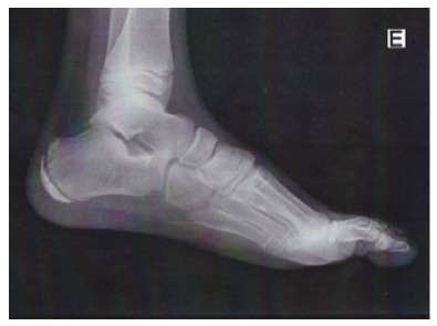
Given the suspicion of overuse pathology, an x-ray and ultrasound of the left foot were performed to evaluate for radiographic signs consistent with the clinical diagnosis of Sever’s disease and exclude other entities such as Achilles tendinitis, retrocalcaneal bursitis, or plantar fasciitis. The radiograph showed mild sclerosis and irregularity of the calcaneal apophysis compatible with calcaneal apophysitis (Figure 1). Ultrasound was normal.
The patient was started on a short course of fixed-dose ibuprofen along with application of topical ice, stretching exercises for the gastrocnemius and soleus muscles, and use of silicone heel cups. He was advised to suspend physical activity until the pain resolved and gradually reintroduce exercise as tolerated.
Significant improvement in symptoms and progressive normalization of gait were noted with treatment. Three weeks later, the patient reported no pain or limitations. He gradually resumed soccer training and had no relapses.
Discussion
The present case illustrates the most common features of Sever’s disease or calcaneal apophysitis, one of the most common causes of heel pain in physically active children.1,3) The clinical presentation of chronic and insidious heel pain that worsens with physical activity and the physical examination findings allowed the clinical diagnosis of Sever’s disease. The radiographic alterations found, including sclerosis and irregularity of the calcaneal apophysis, confirmed the clinical findings. Normal ultrasound allowed exclusion of possible associated complications such as Achilles tendinitis, retrocalcaneal bursitis, and plantar fasciitis. However, since this is a clinical diagnosis, these imaging studies are not necessary for diagnosis.7
Treatment of Sever’s disease includes pharmacologic measures, such as the use of non-steroidal anti-inflammatory drugs, combined with non-pharmacologic measures, such as topical ice application and the use of silicone heel cups and soft-soled running shoes.3,5
Physical activity that aggravates pain should be avoided and resumed only when tolerated.5 Although it may be a potential trigger of heel pain, it does not complicate the disease outcome. On the other hand, physical therapy may be considered, focusing on the plantar fascia and the gastrocnemius and soleus muscles to improve strength and flexibility.1)
Symptoms can sometimes recur and persist for months, disappearing only with the completion of the ossification process. Nevertheless, the importance of adequate adherence to treatment should be emphasized as a key point in controlling associated symptoms.3
Sever’s disease is closely associated with the regular practice of high-impact physical activity, which is the main trigger for symptom onset.3 Avoiding these activities is key to symptomatic control, with progressive reintroduction according to tolerance and gradual reduction of the local inflammatory process.
Conclusion
Sever’s disease is one of the most common causes of calcaneal pain in children, preventing them from performing daily activities. In addition to managing the condition, the family physician plays an active role in reassuring parents about the benignity and favorable prognosis of this entity.
Authorship
Daniela Costa Vieira - Supervision; Writing - original draft; Writing - review & editing
Sara Martins Pinto - Writing - review & editing
Ângela Santos França - Writing - review & editing
Margarida Trigo Silva - Writing - original draft; Writing - review & editing
Claudia Paniagua - Writing - review & editing


