


 Inglés (pdf)
 Articulo en XML
 Referencias del artículo
 Enviar articulo por email
 Citado por SciELO
  Similares en
SciELO
Similares en
SciELO 
 Permalink
In the article by Canakis and Baron entitled “Therapeutic Endoscopic Ultrasound: Current Indications and Future Perspectives” [GE Port J Gastroenterol. 2023, DOI: 10.1159/000529089], Figure 3 was missing from the original publication. Figure 3 is shown here.
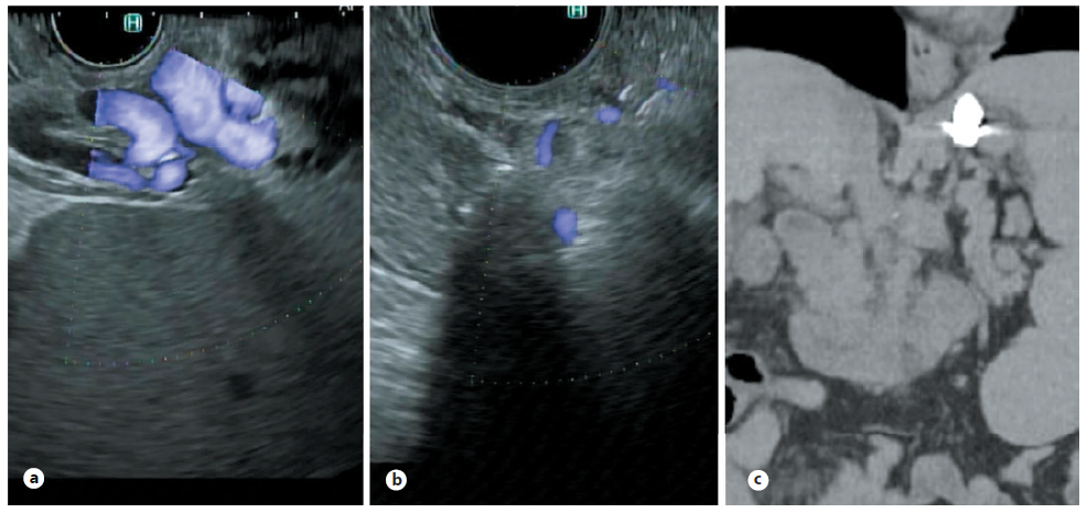
Fig. 3 EUS-guided variceal embolization. a Gastric varices as seen by linear echoendoscope. b Echo image after placement of coils and glue into gastric varix via a 19G needle. Lack of flow as seen by Doppler. c Follow-up coronal CT scan obtained for routine management showing coils in place within the gastric varices

