


 Inglês (pdf)
 Artigo em XML
 Referências do artigo
 Enviar este artigo por email
 Citado por SciELO
  Similares em
SciELO
Similares em
SciELO 
 Permalink
Case Presentation
A 49-year-old woman with history of bicytopenia (anemia and leukopenia) and monoclonal gammopathy (IgG
lambda) not fully clarified presents with portal hypertension and splenomegaly of unclear cause. Abdominal computed tomography (CT) and magnetic resonance imaging (MRI) were performed for further investigation.(Fig. 1)
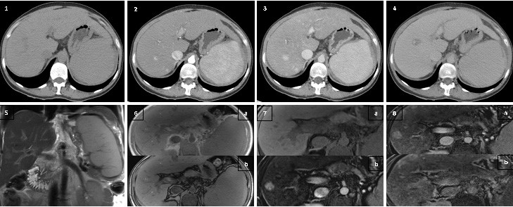
Figures 1: Axial non-enhanced CT; 2. Axial contrast-enhanced CT (hepatic arterial phase); 3. Axial contrast-enhanced CT (portal-venous phase); 4. Axial contrast-enhanced CT (delayed phase); 5. Coronal T2-weighted MRI; 6. Axial T1-weighted gradient echo in-phase (a) and out-of-phase (b) MRI; 7. (a) Axial unenhanced T1-weighted MRI; (b) Axial T1-weighted fat-saturated MRI after enhancement with gadoxetic acid (hepatic arterial phase); 8. (a) Axial T1-weighted fat-saturated MRI after enhancement with gadoxetic acid (portal-venous phase); (b) Axial T1-weighted fat-saturated MRI after enhancement with gadoxetic acid (hepatobiliary phase).

