Serviços Personalizados
Journal
Artigo
Indicadores
-
 Citado por SciELO
 -
 Acessos
Links relacionados
-
 Similares em
SciELO
Compartilhar
GE-Portuguese Journal of Gastroenterology
versão impressa ISSN 2341-4545
GE Port J Gastroenterol vol.24 no.3 Lisboa jun. 2017
https://doi.org/10.1159/000452694
CLINICAL CASE STUDY
Fistulization of J-PEG Jejunal Tube into the Colon in a Patient Treated with Duodopa® Infusion: A Case Report
Fistulização para o Cólon de Sonda Jejunal de PEG-J num Doente Tratado com Infusão de Duodopa®: Caso Clínico
Pedro Russoa, Mariana Costaa, Mário Silvaa, Ary de Sousab, Diana Carvalhoa, Joana Saiotea, Milena Mendesc
Departments of aGastroenterology and bNeurology, Hospital de Santo António dos Capuchos, Centro Hospitalar de Lisboa Central, and cTransplantation Unit, Hospital Curry Cabral, Centro Hospitalar de Lisboa Central, Lisbon, Portugal
* Corresponding author.
ABSTRACT
The continuous delivery of a levodopa/carbidopa gel suspension (Duodopa®) into the small bowel through a jejuna tube inserted via percutaneous endoscopic gastrostomy represents a new treatment method in advanced Parkinson disease. Some severe device-related complications have been described in the last few years. Some of them are associated with phytobezoar formation at the pigtail of the catheter. We present the case of a Parkinson disease patient treated with the Duodopa infusion system complicated by jejunal tube fistulization into the colon. We suggest a possible treatment strategy for this complication, which has not been described in the literature to date.
Keywords: Antiparkinson agents; Parkinson disease; Gastrointestinal endoscopy; Gastrostomy/adverse effects
RESUMO
A perfusão contínua de suspensão em gel de levodopa/carbidopa (Duodopa®) para o intestino delgado através de sonda jejunal, via gastrostomia percutânea endoscópica, representa um novo método de tratamento na Doença de Parkinson avançada. Nos últimos anos têm sido descritas algumas complicações graves, algumas das quais relacionadas com a formação de fitobezoares na ponta pigtail do catéter. Apresentamos o caso de um doente com doença de Parkinson tratado com sistema de infusão Duodopa complicado por fistulização da sonda jejunal para o cólon. Sugere-se uma estratégia terapêutica para esta complicação, não descrita na literatura até à data.
Palavras-Chave: Antiparkinsonianos; Doença de Parkinson; Endoscopia gastrointestinal; Gastrostomia/efeitos adversos
Introduction
The continuous delivery of levodopa/carbidopa gel (Duodopa®) to the small intestine through a jejunal tube in some patients with advanced Parkinson disease (PD) enables less variability in levodopa concentrations, resulting in fewer motor fluctuations and dyskinesia compared with oral administration [1]. The drug-containing gel is steadily pumped into the upper jejunum through an extension tube inserted through a percutaneous endoscopic gastrostomy (J-PEG) [2, 3].
We describe a serious gastrointestinal complication associated with the Duodopa infusion system.
Case Report
A 56-year-old woman with a 26-year history of akinetic rigid PD was proposed Duodopa therapy due to motor symptoms during the previous 7 years, such as fluctuations and dyskinesias, despite optimized oral levodopa/carbidopa treatment. The patient underwent J-PEG for duodenal levodopa/carbidopa gel infusion with a great benefit on parkinsonian symptoms. Two and a half years later, the patient reported worsening of motor symptoms despite increased daily dosage of Duodopa, warranting supplementation with oral levodopa/carbidopa.
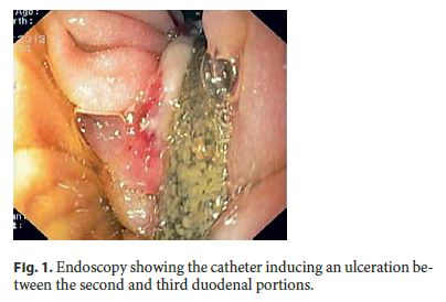
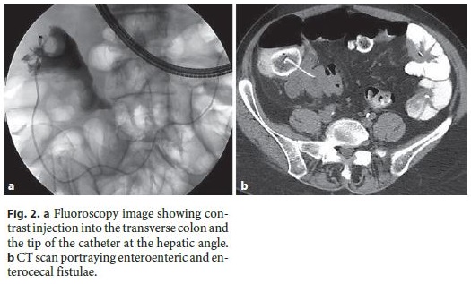
After 3 months, the patient was admitted to our endoscopic department for elective J-PEG substitution. Gastroscopy showed an ulceration area at the transition from the second to the third duodenal portion wall caused by the jejunal tube (Fig. 1). Contrast injection showed the tip of the catheter at the right lower quadrant of the abdomen, apparently in the ascending colon (Fig. 2a). Removal of the jejunal device was impossible because it was blocked when pulled out manually.


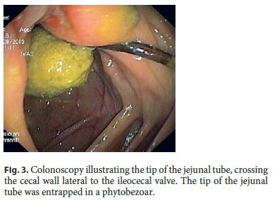
The patient was submitted to a contrast-enhanced CT scan that revealed an intraparietal jejunal tube in the third duodenal portion, with multiple downstream enteroenteric fistulae. The tip of the catheter was placed in the hepatic angle where an irregular parietal thickening was observed (Fig. 2b). Colonoscopy showed the tip of the jejunal tube crossing the cecal wall laterally to the ileocecal valve, entrapped in a phytobezoar (Fig. 3). It was possible to pull the tip of the jejunal tube with an endoscopic snare to the transverse colon, but at this level, the resistance increased considerably due to proximal fixation. After opening the snare, the tip of the catheter spontaneously returned to the ascending colon.

After multidisciplinary discussion (surgery, radiology, neurology, and gastroenterology) regarding the complexity of a surgical solution and the stability of the patients clinical status, a wait-andsee attitude was decided. The patient was maintained on oral levodopa/carbidopa. PEG was removed and the proximal tip of the jejunal tube was cut. Two days after this procedure, complete anal exteriorization of the jejunal tube spontaneously occurred, without any clinical adverse events.
Discussion
Duodopa is a very effective therapy, enabling continuous 24-hour dopaminergic stimulation; therefore, it provides stability in motor and nonmotor symptoms as compared to conventional treatment [4].
Over the last few years, some severe late gastrointestinal complications in patients with Duodopa infusion have been described, many of them related to phytobezoar formation at the tip of the jejunal tube. Klostermann et al. [5] reviewed the medical reports of 52 patients with levodopa/carbidopa gel intestinal infusion. Two patients with mechanical ileus, induced by fiber-rich residuals around the extension tube causing intestinal obstruction, were identified [5]. Similarly, Schrader et al. [6] described 2 cases of phytobezoar formation at the pigtail tip of the jejunal tube, inducing the formation of gastric and jejuna pressure ulcers and jejunal obstruction at the phytobezoar level in 1 of the patients.
Fistulization has also been described as a J-PEG-related complication. Bianco et al. [7] reported a case of a JPEG jejunal tube penetrating through the gastric mucosa, causing fistulization of 3 intestinal loops and jejunal wall perforation. A phytobezoar covering the catheter was seen at the perforation site. The patient required intestinal resection and died from postoperative complications [7]. Olivares et al. [8] reported the case of a patient with mechanical small bowel obstruction. The investigation revealed multiple ulcerations in the duodenum and jejunum and the fistulization of the tube through the jejuna wall into the mesenteric root. The plain abdominal X-ray showed the distal end of the catheter at the ileum level. After an endoscopic attempt to remove the tube, the patient showed signs of pneumoperitoneum. Immediate surgery was required which showed multiple areas of intestinal perforation. The patient died after 2 days [8]. The same authors described a case of a large antro-duodenal ulcer due to the pressure of the jejunal tube. After an endoscopic removal attempt, severe hemorrhage and hemodynamic instability were noted. The pigtail ending of the tube was clogged with a bezoar [8].
Our case illustrates a very serious phytobezoar-related complication of the Duodopa infusion system. Since PEG tubes are frequently used to feed patients who cannot eat solid food, they do not usually predispose to phytobezoars. However, when using J-PEG tubes for intrajejunal Duodopa infusion, patients may ingest a large portion of indigestible fibers and form phytobezoars at the pigtail of the J-PEG tube. Peristaltic movements can propel the bezoar, stretching the J-PEG tube. When the J-PEG tube is stretched tightly, it may carve in the mucosa and cause pressure ulcers [6]. Schrader et al. [6] suggest a low-fiber diet as the probably best way to avoid bezoar formation.
The loss of response with symptom worsening, as in our patient, should alert clinicians to some potentially severe complications (such as tube obstruction, knotting, or fistulization beyond the small bowel) and should suggest prompt endoscopic examination. It is advisable to substitute the jejunal tube every 12 months to avoid late J-PEG tube complications.
Unlike the fatal reports published in the literature, our patient spontaneously expelled the jejunal tube without any adverse events. A wait-and-see attitude should be considered as an option when these complications occur.
References
1 Negreanu L, Popescu BO, Babiuc RD, Ene A, Bajenaru OA, Smarandache GC: Duodopa infusion treatment: a point of view from the gastroenterologist. J Gastrointestin Liver Dis 2011;20:325–327. [ Links ]
2 Nyholm D, Nilsson Remahl IM, Dizdar N, Constantinescu R, Holmberg B, Jansson R, Askmark H: Duodenal levodopa infusion monotherapy vs. oral polypharmacy in advanced Parkinson disease. Neurology 2005;64:216–223. [ Links ]
3 Olanow CW, Obeso JA, Stocchi F: Continuous dopamine-receptor treatment of Parkinsons disease: scientific rationale and clinical implications. Lancet Neurol 2006;5:677–687. [ Links ]
4 Nyholm D, Askmark H, Gomes-Trolin C, Knutson T, Lennernäs H, Nyström C, et al: Optimizing levodopa pharmacokinetics: intestinal infusion versus oral sustained-release tablets. Clin Neuropharmacol 2003;26:156–163. [ Links ]
5 Klostermann F, Jugel C, Bömelburg M, Marzinzik F, Ebersbach G, Müller T: Severe gastrointestinal complications in patients with levodopa/carbidopa intestinal gel infusion. Mov Disord 2012;27:1704–1705. [ Links ]
6 Schrader C, Böselt S, Wedemeyer J, Dressler D, Weismüller TJ: Asparagus and jejunalthrough-PEG: an unhappy encounter in intrajejunal levodopa infusion therapy. Parkinsonism Relat Disord 2010;17:67–69. [ Links ]
7 Bianco G, Vuolo G, Ulivelli M, Bartalini S, Chieca R, Rossi A, et al: A clinically silent, but severe, duodenal complication of duodopa infusion. J Neurol Neurosurg Psychiatry 2012;83:668–670. [ Links ]
8 Olivares A, Collado D, Muñoz-Navas M, Calvo M, Olivo E, Chico I, et al: Complications of percutaneous endoscopic gastrostomy-jejunostomy for levodopa/carbidopa infusion in advanced Parkinsons disease. Gastroenterology Insights 2012;4:13–15. [ Links ]
Statement of Ethics
This study did not require informed consent or review/approval by the appropriate ethics committee.
Disclosure Statement
The authors have no conflicts of interest to disclose.
* Corresponding author.
Dr. Pedro Russo
Department of Gastroenterology, Hospital de Santo António dos Capuchos
Alameda de Santo António dos Capuchos
PT–1169–050 Lisbon (Portugal)
E-Mail pedro.me.russo@gmail.com
Received: August 8, 2016; Accepted after revision: September 26, 2016


