


Services on Demand
Journal
Article
 English (pdf)
 Article in xml format
 Article references
 Send this article by e-mail
Indicators
-
 Cited by SciELO
 -
 Access statistics
Related links
-
 Similars in
SciELO
Similars in
SciELO
Share

 Permalink
Angiologia e Cirurgia Vascular
Print version ISSN 1646-706X
Angiol Cir Vasc vol.9 no.3 Lisboa Sept. 2013
VASCULAR IMAGES
Bilateral isolated aneurysms of profunda femoris artery
Aneurismas isolados da artéria femoral profunda
José Tiagoa,*, Pedro Martinsa, Viviana Manuela, Carlos Martinsa, José Fernandes e Fernandesa
aDepartment of Angiology and Vascular Surgery, Hospital Universitário de Santa Maria, Lisbon, Portugal
Introduction
First described by Pappas et al.1 arteriosclerotic aneurysms of profunda femoris are very rare entities.1-5 Because of its anatomical position in the muscular fascia and wall characteristics the profunda artery is less affected by aneurismal disease than other peripheral arteries and true aneurysms account for less than 0.5%2 of all peripheral aneurysms and 1-6,6%3,4 of all femoral arteries aneurysms.
Bilateral profunda femoris artery aneurysms (BPFAA) are even rarer and without a context of polianeurysmatic disease few cases are described in literature.5
Case report
A 72 years old man presented with complaints of acute swelling and pulsatile mass in the proximal left thigh (fig. 1). On past medical history the patient was a former smoker, had a bilateral inguinal hernia repair, open prostatectomy for benign prostatic hyperplasia and was being treated for arterial hypertension and atrial fibrillation. He denied other comorbidities, history of trauma or needle use in the left groin.
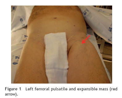
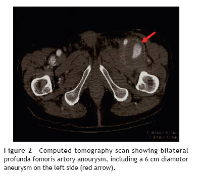
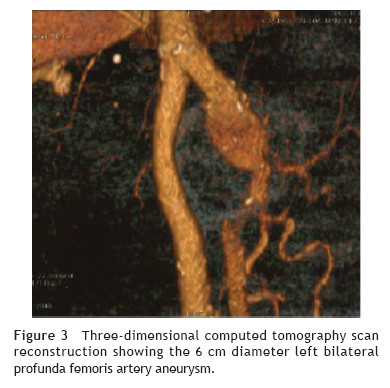
On physical examination there was a large pulsatile and expansible mass in the left medial thigh region. The duplex scan examination showed BPFAA with a large left profunda femoris artery aneurysm (PFAA) CT scan revealed a 6 cm diameter PFAA in the left tight and a right 1.7 cm PFAA (Figs. 2 and 3). No other aneurysms were present at the CT screening.
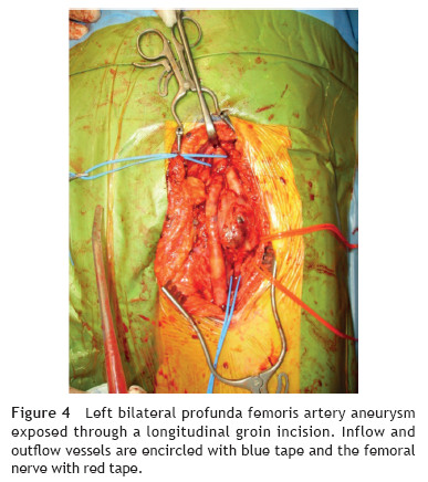
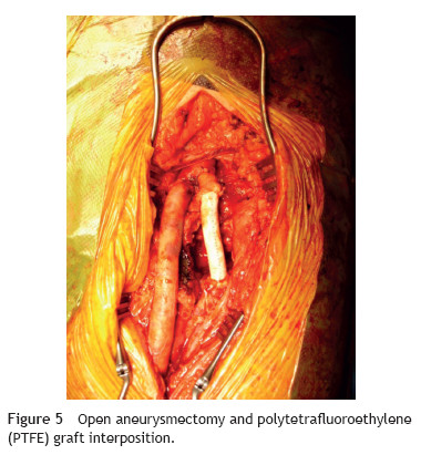
The patient underwent successful surgical repair with open aneurysmectomyand graft repair with polytetra fluoroethylene (PTFE) interposition on the left side. The right PFAA was maintained in close surveillance (Figs. 4 and 5).
Conclusion
Isolated BPFAA are extremely rare and asymptomatic most of the time, when symptoms are present, surgical repair is prompt recommended to prevent complications such as femoral nerve and veins compression and aneurysm rupture with great risk of limb loss. Treatment of PFAA is safe maintains profunda artery function, eliminates the presence of compressive neurologic signs and reduces deep venous thrombosis risk.
Bibliografia
1. Pappas G, Janes J, Bernatz P, Schriger A. Femoral aneurysms. JAMA. 1964;190:489–93. [ Links ]
2. Rich PB, Wolk SW, Sarosi MJ, Shanley CJ. Endovascular management of a true aneurysm of the profunda femoris artery: a case report. Vasc Endovascular Surg. 2000;34:467–70. [ Links ]
3. Posner SR,Wilensky J, Dimick J, Henke PK. A true aneurysm of the profunda femoris artery: a case report and review of the English language literature. Ann Vasc Surg. 2004;18:740–6. [ Links ]
4. Cutler BS, Darling RC. Surgical management of arteriosclerotic femoral aneurysms. Surgery. 1973;74:764–73. [ Links ]
5. Gemayel G, Mugnai D, Khabiri E, Sierra J, Murith N, Kalangos A. Isolated bilateral profunda femoris artery aneurysm. Ann Vasc Surg. 2010;24:824.e11–824.e13. [ Links ]
E-mail address: josetiagus@gmail.com
Recebido a 1 de maio de 2013;
Aceite a 15 de junho de 2013

