Inglês (pdf)
 Artigo em XML
 Referências do artigo
 Enviar este artigo por email
 Citado por SciELO
  Similares em
SciELO
Similares em
SciELO 
 Permalink
A 12 month-old male infant was referred to the paediatric pulmonary unit of our tertiary hospital for further investigation.
At the age of 4 months, he had been referred by his family doctor to a secondary hospital because of respiratory distress since the first month of life and failure to thrive. At that time, clinical observation revealed an increase in the anteroposterior diameter of the chest, elevated respiratory rate, and subcostal and supraclavicular retractions. On auscultation, crackles were heard in both lung bases without wheezing. Peripheral blood oxygen saturation was 92% in room air. Laboratory investigations, which included alpha-1 antitrypsin levels and sweat test, were within in the normal range. Chest x-ray (not shown) revealed bilateral lung hyperinflation and no further remarks. He was treated with supplemental oxygen and inhaled corticoids with mild clinical improvement.
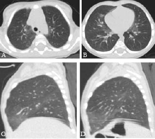
A high-resolution chest computed tomography (CT) was performed at our department (figure 1). What is your diagnosis?