


 Inglês (pdf)
 Artigo em XML
 Referências do artigo
 Enviar este artigo por email
 Citado por SciELO
  Similares em
SciELO
Similares em
SciELO 
 Permalink
Introduction
Bowel intussusception, a process in which a proximal segment of intestine invaginates into the lumen of the adjacent intestinal, is a rare entity which occurs in 2-3 cases/1 000 000 per year and causes 1%-5% of bowel obstruction cases.1,2 This entity appears more frequently in children and males and is the second most common cause of acute abdomen in children.1,3
Classification of intussusception can be done according to its etiology or location. In the adult population only 8%-20% of cases are idiopathic/primary.4,5 Several conditions like benign polyp, lipoma, diverticulum, malignant tumors, HIV infection, celiac sprue or intramural hematoma may predispose to intussusception.2,3 Intussusception can be categorized according to the involved bowel segment: 1) entero-enteric; 2) colo-colic; 3) ileo-colic; and 4) ileo-cecal.6 A literature review from Honjo et al has revealed a malignant tumor etiology in 48% of patients with colo-colic intussusception and in 17% of those with entero-enteric.7 The most frequent histologic findings are adenocarcinoma or metastatic melanoma, respectively.1,8 Wang et al, in his series of 292 cases of intussusception, found that the most common enteric etiology were lipomas and not metastatic lesions.9
Unlike children, the majority of adult patients with intussusception present nonspecific symptoms such as dull abdominal pain, changes in bowel habits and abdominal distension. The classic triad of abdominal pain, vomiting and lower gastrointestinal bleeding rarely occurs.1,3,10 Acute symptoms due to complete bowel obstruction appear in less than 20% of the patients.7 In few cases, patients may develop sepsis and bowel perforation or necrosis.3 Physicians should also inquire for constitutional symptoms, such as weight loss, fatigue or anorexia.8 Despite the nonspecificity of the condition, detection of adult intussusception has increased with the widespread use of novel diagnostic imaging techniques.
Case Report
A 44-year-old male patient presented to the urgency department with intermittent colicky epigastric pain since the day before, which initiated in the peri-umbilical area and was associated with bilious vomiting. He denied any other symptoms, namely fever, abdominal distension, digestive hemorrhage or change of bowel habits.
His past medical and family history was irrelevant, and no previous endoscopic studies had been performed. His vital signs were within reference values. Physical examination revealed dehydration, abdominal distension, epigastrium tenderness and no palpable masses. On rectal examination, no masses or signs of bleeding were found. Adequate hydro-electrolyte resuscitation was initiated on admission.
Initial laboratory data showed white cell count of 12.59 x 10⁹/L, with 90% neutrophils and a C-reactive protein (CRP) of 8.10 mg/L. Standing plain abdominal radiography revealed dilation of small bowel with gas-fluid levels (Fig. 1). There were no signs of perforation. Contrast-enhanced computed tomography (CT) scan of abdomen and pelvis showed a small bowel obstruction and a typical “sausage” sign near the ileocecal junction surrounded by its mesenteric fat, suggestive of ileoileal intussusception (Fig. 2). There were no indirect signs of a malignant tumor.

Figure 1 Abdominal radiograph on admission: Lower abdomen axial CT scan: small bowel intussusception with dilation of the edematous upstream bowel loops and “sausage”-shape soft tissue mass.
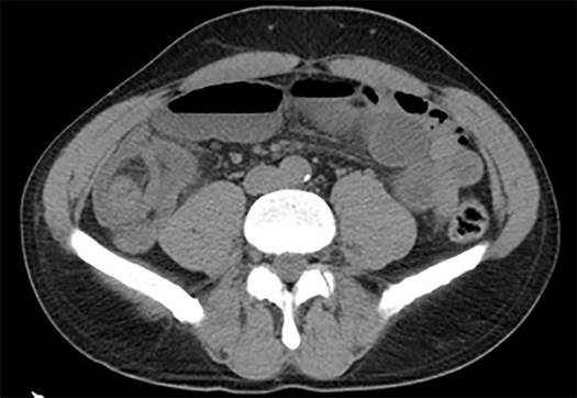
Figure 2 Abdominal CT on admission: small bowel intussusception with dilation of the edematous upstream bowel loops and “sausage”-shape soft tissue mass.
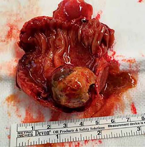
The patient was taken to the operating room for a laparotomy. On exploration, we found a grossly dilated small bowel and a 2.5 x 3 cm palpable intraluminal lump, already without intussusception. This mass was soft, mobile, with no adherence to the mesentery. Also, no macroscopic signs of local invasion or metastatic lymph nodes were found. We resected the involved segment of the bowel with a 3 cm margin on either side, removed the closest lymph nodes and performed a primary anastomosis. Cut section of resected specimen showed a pedunculated polyp with intact overlying mucosa (Fig. 3). The histopathology report revealed a 2.7 x 2.3 x 3 cm submucosal lipoma of the terminal ileum. There was no evidence of dysplasia or malignancy.
Post-operative period was uneventful, and the patient was discharged on the fifth postoperative day.
Discussion
Any lesion in the bowel that alters normal peristaltic activity can initiate an invagination. In this case, the causing lesion was a lipoma, a benign tumor of mesenchymal origin, usually solitary, that can be found anywhere along the gastrointestinal tract, most commonly in the colon.10,11 They can cause of acute intestinal obstruction as presented above, but also nonspecific gastrointestinal symptoms or blood loss. During the first decade of this century, only 51 cases of adult intussusception caused by lipomas were reported in the literature.10 During the second decade, only 55 cases were published as case reports in PubMed (pubmed.ncbi.nlm.nih.gov/), mainly in the colon.
Lipomas account for 5% of all gastrointestinal tumors and are the second most common benign tumor of the small bowel.2,11 The peak age of incidence is between 50 and 70 years old and it appears to affect more women than men.1,8,12 They are usually asymptomatic, especially if they are small. Symptoms are usually vague, such as abdominal pain, diarrhea or vomiting. A palpable mass is present in fewer than 40% of the cases.11 When larger than 2 cm, they might ulcerate and cause acute or chronic anemia due to blood losses.1,11 Due to their intramural location, lipomas may also be one of the causes of intussusception, and therefore of acute intestinal obstruction. Our patient presented with acute obstruction rather than nonspecific symptoms, which relates to its intramural location and large size of the lesion.
The variability in clinical presentations often requires the use of imaging methods.8 CT scan is the method of choice for the diagnosis of intussusception, with a reported sensitivity of more than 70% and 100% of specificity.1,3,9,10 Classic imaging signs consist of “target” or “sausage” signs, a mass lesion with Hounsfield units between -80 and -120 and a crescent-like fatty mass.3,5,11 In our case report, a typical “sausage” sign led us to consider intussusception as the most probable diagnostic. Computed tomography (CT) scan can also provide indirect signs of bowel ischemia (gas in the bowel wall, in the mesenteric and portal veins, and mesenteric fat stranding) or malignancy (larger size and thickness of the mass, intratumoral calcification/hyperattenuation, regional lymphadenopathy, direct invasion of adjacent organs or metastasis).1,13 However, due to economic and safety reasons, ultrasound is usually the first tool in the diagnosis, which is more appropriate if a palpable mass is found. An echogenic mass with pseudokidney sign, a trident sign or a doughnut sign can make the diagnosis of intussusception on ultrasound.1,2 Other approaches for the diagnosis of intussusception are endoscopy, plain abdominal radiography, barium studies, endoscopy, colonoscopy or magnetic resonance imaging.3,4
Unlike children, adults’ intussusception rarely resolves with barium studies due to the high incidence of underlying anatomical abnormalities, and surgical treatment is usually indicated.5,9 The extension of surgery depends on the site, cause and degree of obstruction. Resection with or without prior reduction is a controversial issue because reduction of an undiagnosed lesion could lead to transperitoneal or vascular seeding if an underlying malignant lesion is the cause. It can also increase the risk of bowel perforation and venous embolization.7,14 Therefore, reduction of the intussusception should be reserved for patients in whom resection may cause short gut syndrome or in whom benign diagnosis has been made previously.10,15 Intraoperative colonoscopy may help to distinguish benign from malignant lesions before reduction.9 Irreversible bowel ischemia or marked inflammation are contraindications for reduction.1,7,10 For acute intussusception, an en bloc resection without previous reduction is the recommended treatment.2,10 For lesions on the right side of the colon, resection with primary anastomosis is preferred. For left-sided lesions, it is safer to perform resection with a colostomy and Hartmann’s pouch, with re-anastomosis at a second stage.9
Some authors recommend operative reduction for small bowel before resection with primary anastomosis, once again due to concerns of a short gut syndrome and because small bowel lesions were malignant in less than half of the cases (12%-40%)5,7,9 Laparoscopic approach has been reported as a possibility based on the clinical condition of the patient, the location and extent of intussusception and surgeons’ expertise. In our case, we chose to perform an open approach due to exuberant dilation of the small bowel. An ileoileal intussusception was diagnosed intraoperatively without any macroscopic malignancy criteria, therefore we performed a resection of the bowel involving the lesion, after spontaneous reduction. Endoscopic resection has been described, although complications such as perforation or hemorrhage have been reported.3
The major risk factor affecting intussusception prognosis is the cause of the lesion, with overall mortality rates ranging between 8.7% for benign lesions to 52.4% with malignant.3,4 Lipomas are a benign condition with no reports of malignant transformation and therefore with good prognosis.1 There is no report of mortality due to lipoma in patients successfully treated.14

