


Servicios Personalizados
Revista
Articulo
 Inglés (pdf)
 Articulo en XML
 Referencias del artículo
 Enviar articulo por email
Indicadores
-
 Citado por SciELO
 -
 Accesos
Links relacionados
-
 Similares en
SciELO
Similares en
SciELO
Compartir

 Permalink
GE-Portuguese Journal of Gastroenterology
versión impresa ISSN 2341-4545
GE Port J Gastroenterol vol.27 no.1 Lisboa feb. 2020
https://doi.org/10.1159/000500209
ENDOSCOPIC SNAPSHOT
An Unusual Cause of Cholangitis
Uma causa incomum de colangite
Pablo Cortegoso Valdiviaa, Ludovica Veneziaa, Stefano Rizzaa, Luigi Chiusab, Claudio Giovanni De Angelisc
aGastroenterology Unit, University of Turin, Turin, Italy; bPathology Unit, University of Turin, AOU Città della Salute e della Scienza, Turin, Italy; cGastroenterology Unit, AOU Città della Salute e della Scienza, Turin, Italy
* Corresponding author.
Keywords: Cholangioscopy, Cholestasis, Diagnosis, Endoscopic ultrasonography, Endoscopy, Endosonography, Liver transplantation
Palavras-chave: Colangioscopia, Colestase,Diagnóstico, Ecoendoscopia, Transplante hepático
A 57-year-old man with unremarkable previous medical history presented with acute cholangitis and cholestasis. An abdominal computed tomography showed a dilation of the biliary system with hypodense irregular filling defects in the common bile duct (CBD) and previously unknown liver cirrhosis.
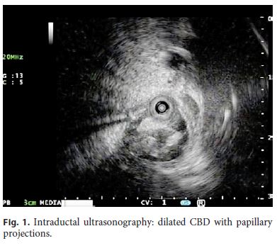
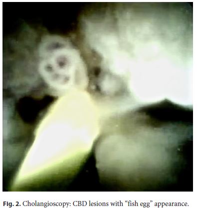
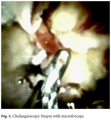
The papilla was normal; a subsequent intraductal ultrasonography showed multiple papillary projections in the CBD (Fig. 1). These findings were confirmed by cholangioscopy [1], which identified multiple lesions with “fish-egg” appearance protruding in a dilated CBD filled with whitish mucus (Fig. 2). Biopsies with microforceps on the papillary lesions (Fig. 3) were performed, and histological examination highlighted the presence of papillary proliferation with focal high-grade dysplasia without stromal invasion, thus confirming the final diagnosis of intraductal papillary neoplasm BillN-3 (Fig. 4) [2].
Due to the high rate of malignant transformation of this disease despite its slow progression, after multidisciplinary discussion the patient has now been referred and selected for orthotopic liver transplantation [3].
References
1 Parsi MA. Biliary papillomatosis: diagnosis with direct peroral cholangioscopy. Gastrointest Endosc. 2015 Jan;81(1):231–2. [ Links ]
2 Wan XS, Xu YY, Qian JY, Yang XB, Wang AQ, He L, et al. Intraductal papillary neoplasm of the bile duct. World J Gastroenterol. 2013 Dec;19(46):8595–604. [ Links ]
3 Luvira V, Pugkhem A, Bhudhisawasdi V, Uttaravichien TU, Sripanuskul A, Pongskul J, et al. Papillomatosis of the Biliary Tree and Gallbladder: Successful Treatment With Repeated Resection and Liver Transplant. Exp Clin Transplant. 2017 Dec. doi: 10.6002/ect.2017.0121. [ Links ]
Statement of Ethics
This material has not been published in whole or in part elsewhere; the manuscript is not currently being considered for publication in another journal; all authors have been personally and actively involved in substantive work leading to the manuscript and will hold themselves jointly and individually responsible for its content.
Disclosure Statement
All authors declare that there is no conflict of interest related to this manuscript.
* Corresponding author.
Pablo Cortegoso Valdivia
Gastroenterology Unit, University of Turin
Corso Bramante 88–90
IT–10126 Turin (Italy)
E-Mail cortegosopablo@yahoo.it
Received: February 28, 2019; Accepted after revision: March 29, 2019
Author Contributions
P. Cortegoso Valdivia and L. Venezia equally contributed in writing the manuscript, S. Rizza contributed to its editing and selection of the images, L. Chiusa analyzed and provided images of the histological specimens, and C.G. De Angelis contributed with critical revision.

